9QGG | pdb_00009qgg

Consensus structure of dUBA1-UbDha-dBIRC6 trapped ternary complex


Experimental Data Snapshot

  • Method: ELECTRON MICROSCOPY
  • Resolution: 2.58 Å
  • Aggregation State: PARTICLE 
  • Reconstruction Method: SINGLE PARTICLE 

wwPDB Validation   3D Report Full Report


This is version 1.1 of the entry. See complete history


Literature

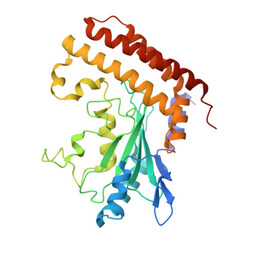
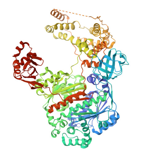
UBA6 specificity for ubiquitin E2 conjugating enzymes reveals a priority mechanism of BIRC6.

Riechmann, C.Ellison, C.J.Anderson, J.W.Hofmann, K.Sarkies, P.Elliott, P.R.

(2025) Nat Struct Mol Biol 

  • DOI: https://doi.org/10.1038/s41594-025-01717-z
  • Primary Citation of Related Structures:  
    9QGG, 9QGI, 9QGR, 9QGW, 9QH5, 9QHI, 9QIA, 9QIC, 9QIG, 9QII, 9QIM, 9QIO, 9QIP, 9QIV

  • PubMed Abstract: 

    In mammals, ubiquitylation is orchestrated by the canonical ubiquitin-activating E1 enzyme UBA1 and the orthogonal E1 UBA6. Growing evidence underscores the essentiality of both E1s, which differentiate between 29 active ubiquitin-conjugating enzymes (E2s). The mechanisms governing this distinction have remained unclear. Here we establish a framework for ubiquitin E1-E2 specificity. Focusing on UBA6-controlled ubiquitylation cascades, we reveal that BIRC6, a UBA6-exclusive E2, gains priority over all other UBA6-competent E2s, underpinning the functional importance of defined UBA6-BIRC6 ubiquitylation events in regulating cell death, embryogenesis and autophagy. By capturing BIRC6 receiving ubiquitin from UBA6 in different states, we observe BIRC6 engaging with the UBA6 ubiquitin fold domain, driving an exceptionally high-affinity interaction that is modulated by the UBA6 Cys-Cap loop. Using this interaction as a template, we demonstrate how to confer activity between E2s and their noncognate E1, providing a tool to delineate E1-E2-dependent pathways. Lastly, we explain how BIRC6 priority does not lead to inhibition of UBA6, through a bespoke thioester switch mechanism that disengages BIRC6 upon receiving ubiquitin. Our findings propose a concept of hierarchy of E2 activity with cognate E1s, which may explain how ubiquitin E1s can each function with over a dozen E2s and orchestrate E2-specific cellular functions.


  • Organizational Affiliation
    • Department of Biochemistry, University of Oxford, Oxford, UK.

Macromolecules
Find similar proteins by:  (by identity cutoff)  |  3D Structure
Entity ID: 1
MoleculeChains Sequence LengthOrganismDetailsImage
BIR repeat containing ubiquitin-conjugating enzyme, isoform B305Drosophila melanogasterMutation(s): 0 
Gene Names: BruceBRUCEdBRUCEDmel\CG6303mod86CG6303Dmel_CG6303
EC: 2.3.2.23
UniProt
Find proteins for A0A0B4KG50 (Drosophila melanogaster)
Explore A0A0B4KG50 
Go to UniProtKB:  A0A0B4KG50
Entity Groups  
Sequence Clusters30% Identity50% Identity70% Identity90% Identity95% Identity100% Identity
UniProt GroupA0A0B4KG50
Sequence Annotations
Expand
  • Reference Sequence
Find similar proteins by:  (by identity cutoff)  |  3D Structure
Entity ID: 2
MoleculeChains Sequence LengthOrganismDetailsImage
E1 ubiquitin-activating enzyme1,010Drosophila melanogasterMutation(s): 0 
Gene Names: 
EC: 6.2.1.45
UniProt
Find proteins for Q8T0L3 (Drosophila melanogaster)
Explore Q8T0L3 
Go to UniProtKB:  Q8T0L3
Entity Groups  
Sequence Clusters30% Identity50% Identity70% Identity90% Identity95% Identity100% Identity
UniProt GroupQ8T0L3
Sequence Annotations
Expand
  • Reference Sequence
Find similar proteins by:  (by identity cutoff)  |  3D Structure
Entity ID: 3
MoleculeChains Sequence LengthOrganismDetailsImage
Polyubiquitin-CC,
D [auth E]
76Homo sapiensMutation(s): 1 
Gene Names: UBC
UniProt & NIH Common Fund Data Resources
Find proteins for P0CG48 (Homo sapiens)
Explore P0CG48 
Go to UniProtKB:  P0CG48
PHAROS:  P0CG48
GTEx:  ENSG00000150991 
Entity Groups  
Sequence Clusters30% Identity50% Identity70% Identity90% Identity95% Identity100% Identity
UniProt GroupP0CG48
Sequence Annotations
Expand
  • Reference Sequence
Small Molecules
Ligands 1 Unique
IDChains Name / Formula / InChI Key2D Diagram3D Interactions
AMP
Query on AMP

Download Ideal Coordinates CCD File 
E
ADENOSINE MONOPHOSPHATE
C10 H14 N5 O7 P
UDMBCSSLTHHNCD-KQYNXXCUSA-N
Experimental Data & Validation

Experimental Data

  • Method: ELECTRON MICROSCOPY
  • Resolution: 2.58 Å
  • Aggregation State: PARTICLE 
  • Reconstruction Method: SINGLE PARTICLE 

Structure Validation

View Full Validation Report


Entry History & Funding Information

Deposition Data


Funding OrganizationLocationGrant Number
UK Research and Innovation (UKRI)United KingdomBB/Y008936/1
Cancer Research UKUnited KingdomDRCPFA-Jun24/100003

Revision History  (Full details and data files)

  • Version 1.0: 2025-10-29
    Type: Initial release
  • Version 1.1: 2025-12-17
    Changes: Data collection, Database references