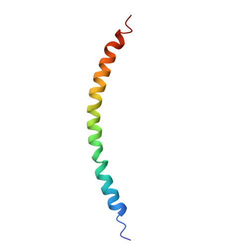
Structure of the virulence-associated Neisseria meningitidis filamentous bacteriophage MDA Phi.
Bohning, J., Graham, M., Coureuil, M., Tarafder, A.K., Meyer, J., Nassif, X., Bille, E., Bharat, T.A.M.(2025) Proc Natl Acad Sci U S A 122: e2420157122-e2420157122
- PubMed: 40540604 Search on PubMedSearch on PubMed Central
- DOI: https://doi.org/10.1073/pnas.2420157122
- Primary Citation Related Structures:
9QG9 - PubMed Abstract:
Neisseria meningitidis is a human commensal bacterium that can opportunistically invade the bloodstream and cross the blood-brain barrier, where it can cause septicemia and meningitis. These diseases, if left untreated, can be lethal within hours. Hyperinvasive N. meningitidis strains often express a genomically encoded filamentous bacteriophage called MDAΦ, which promotes colonization of mucosal host surfaces to facilitate bacterial invasion. How this phage is organized and how it promotes biofilm formation and infection at the molecular level is unclear. Here, we present an electron cryomicroscopy structure of the MDA phage, showing that MDAΦ is a class I filamentous inovirus, with the major capsid protein (MCP) arranged within the phage as a highly curved and densely packed α-helix. Comparison with other filamentous bacteriophages offers clues about inoviral genome encapsidation mechanisms, providing a framework for understanding the evolutionary diversity of inoviruses. A disordered, N-terminal segment in the MCP presents hydrophobic patches on the surface of assembled phage particles, which, together with electron cryotomography data of phage bundles, furnishes a structural rationale for phage-phage interactions that were seen previously in an epithelium adhesion infection model of N. meningitidis . Taken together, our results shed light on the structure, organization, and higher-order assembly of a biomedically relevant phage encoded in the genome of a human pathogen. Molecular insights gleaned from this study increase our understanding of phage evolution, phage-mediated bacterial adhesion, and pathogenicity.
- Structural Studies Division, Medical Research Council Laboratory of Molecular Biology, Cambridge CB2 0QH, United Kingdom.
Organizational Affiliation: