9QBG | pdb_00009qbg

HER2/ErbB2 extracellular domain (ECD) in extended conformation in complex with trastuzumab (TZB) antibody


Experimental Data Snapshot

  • Method: ELECTRON MICROSCOPY
  • Resolution: 3.60 Å
  • Aggregation State: PARTICLE 
  • Reconstruction Method: SINGLE PARTICLE 

Starting Model: experimental
View more details

wwPDB Validation   3D Report Full Report


This is version 1.0 of the entry. See complete history


Literature

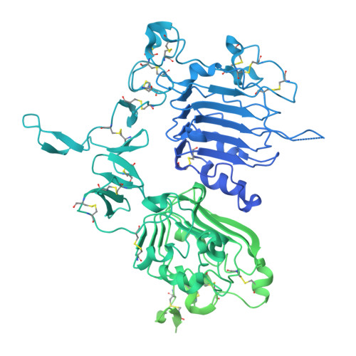
Structural analysis of HER2-trastuzumab complex reveals receptor conformational adaptation.

Vacca, S.Gragera, M.Buschiazzo, A.Herreros, D.Krieger, J.M.Bonn-Garcia, S.Melero, R.Sorzano, C.O.Carazo, J.M.Medalia, O.Pluckthun, A.

(2025) Sci Adv 11: eadu9945-eadu9945

  • DOI: https://doi.org/10.1126/sciadv.adu9945
  • Primary Citation of Related Structures:  
    9QBF, 9QBG, 9QBH

  • PubMed Abstract: 

    Human epidermal growth factor receptor-2 (HER2) is a receptor tyrosine kinase, associated with a variety of malignant tumors, usually through overexpression, resulting in aberrant signaling. Trastuzumab (TZB), one of the monoclonal antibodies (mAbs) used in combination with chemotherapy, has become a major therapeutic for HER2-overexpressing cancers. Current structural understanding of HER2 and its interactions with other receptors and with different affinity agents has relied on numerous structures of individual domains of HER2. Here, we subjected purified near full-length HER2 to single-particle cryo-electron microscopy (cryo-EM) analysis. Besides the canonical conformation described in previous structural studies, we report a previously unreported conformation of the HER2 extracellular domain that is stabilized upon TZB binding, which might hamper association with HER3, a receptor with which HER2 forms an oncogenic unit. Together, our findings provide insights into the conformational dynamics of the HER2 receptor and the mechanism of action of TZB.


  • Organizational Affiliation
    • Department of Biochemistry, University of Zürich, Winterthurerstrasse 190, CH-8057 Zürich, Switzerland.

Macromolecules
Find similar proteins by:  (by identity cutoff)  |  3D Structure
Entity ID: 1
MoleculeChains Sequence LengthOrganismDetailsImage
Receptor tyrosine-protein kinase erbB-2,Green fluorescent protein1,311Homo sapiensAequorea victoria
This entity is chimeric
Mutation(s): 3 
Gene Names: ERBB2HER2MLN19NEUNGLGFP
EC: 2.7.10.1
UniProt & NIH Common Fund Data Resources
Find proteins for P04626 (Homo sapiens)
Explore P04626 
Go to UniProtKB:  P04626
PHAROS:  P04626
GTEx:  ENSG00000141736 
Entity Groups  
Sequence Clusters30% Identity50% Identity70% Identity90% Identity95% Identity100% Identity
UniProt GroupP04626
Sequence Annotations
Expand
  • Reference Sequence
Experimental Data & Validation

Experimental Data

  • Method: ELECTRON MICROSCOPY
  • Resolution: 3.60 Å
  • Aggregation State: PARTICLE 
  • Reconstruction Method: SINGLE PARTICLE 
EM Software:
TaskSoftware PackageVersion
MODEL REFINEMENTCoot0.9.6
MODEL REFINEMENTPHENIX1.21.1_5286:
MODEL REFINEMENTRosettaEM2024.09+release.06b3cf8ad0

Structure Validation

View Full Validation Report


Entry History & Funding Information

Deposition Data


Funding OrganizationLocationGrant Number
European Research Council (ERC)European UnionHighResCells (ERC-2018-SyG, Proposal 810057)

Revision History  (Full details and data files)

  • Version 1.0: 2025-08-06
    Type: Initial release