9PZL | pdb_00009pzl

Pyruvate phosphate dikinase in complex with AMP-PNP and sulfate ions


Experimental Data Snapshot

  • Method: X-RAY DIFFRACTION
  • Resolution: 3.00 Å
  • R-Value Free: 
    0.302 (Depositor), 0.314 (DCC) 
  • R-Value Work: 
    0.219 (Depositor), 0.229 (DCC) 
  • R-Value Observed: 
    0.225 (Depositor) 

Starting Model: experimental
View more details

wwPDB Validation   3D Report Full Report


Ligand Structure Quality Assessment 


This is version 1.0 of the entry. See complete history


Literature

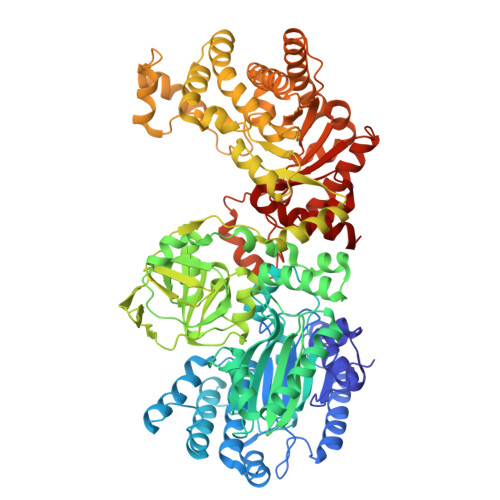
Swiveling domain mechanism in pyruvate phosphate dikinase.

Lim, K.Read, R.J.Chen, C.C.Tempczyk, A.Wei, M.Ye, D.Wu, C.Dunaway-Mariano, D.Herzberg, O.

(2007) Biochemistry 46: 14845-14853

  • DOI: https://doi.org/10.1021/bi701848w
  • Primary Citation of Related Structures:  
    2R82, 9PZL

  • PubMed Abstract: 

    Pyruvate phosphate dikinase (PPDK) catalyzes the reversible conversion of phosphoenolpyruvate (PEP), AMP, and Pi to pyruvate and ATP. The enzyme contains two remotely located reaction centers: the nucleotide partial reaction takes place at the N-terminal domain, and the PEP/pyruvate partial reaction takes place at the C-terminal domain. A central domain, tethered to the N- and C-terminal domains by two closely associated linkers, contains a phosphorylatable histidine residue (His455). The molecular architecture suggests a swiveling domain mechanism that shuttles a phosphoryl group between the two reaction centers. In an early structure of PPDK from Clostridium symbiosum, the His445-containing domain (His domain) was positioned close to the nucleotide binding domain and did not contact the PEP/pyruvate-binding domain. Here, we present the crystal structure of a second conformational state of C. symbiosum PPDK with the His domain adjacent to the PEP-binding domain. The structure was obtained by producing a three-residue mutant protein (R219E/E271R/S262D) that introduces repulsion between the His and nucleotide-binding domains but preserves viable interactions with the PEP/pyruvate-binding domain. Accordingly, the mutant enzyme is competent in catalyzing the PEP/pyruvate half-reaction but the overall activity is abolished. The new structure confirms the swivel motion of the His domain. In addition, upon detachment from the His domain, the two nucleotide-binding subdomains undergo a hinge motion that opens the active-site cleft. A similar hinge motion is expected to accompany nucleotide binding (cleft closure) and release (cleft opening). A model of the coupled swivel and cleft opening motions was generated by interpolation between two end conformations, each with His455 positioned for phosphoryl group transfer from/to one of the substrates. The trajectory of the His domain avoids major clashes with the partner domains while preserving the association of the two linker segments.


  • Organizational Affiliation
    • Center for Advanced Research in Biotechnology, University of Maryland Biotechnology Institute, Rockville, Maryland 20850, USA.

Macromolecules
Find similar proteins by:  (by identity cutoff)  |  3D Structure
Entity ID: 1
MoleculeChains Sequence LengthOrganismDetailsImage
Pyruvate, phosphate dikinase
A, B, C, D
874[Clostridium] symbiosumMutation(s): 0 
Gene Names: ppdK
EC: 2.7.9.1
UniProt
Find proteins for P22983 (Clostridium symbiosum)
Explore P22983 
Go to UniProtKB:  P22983
Entity Groups  
Sequence Clusters30% Identity50% Identity70% Identity90% Identity95% Identity100% Identity
UniProt GroupP22983
Sequence Annotations
Expand
  • Reference Sequence
Small Molecules
Ligands 2 Unique
IDChains Name / Formula / InChI Key2D Diagram3D Interactions
ANP (Subject of Investigation/LOI)
Query on ANP

Download Ideal Coordinates CCD File 
E [auth A],
I [auth B],
M [auth C],
Q [auth D]
PHOSPHOAMINOPHOSPHONIC ACID-ADENYLATE ESTER
C10 H17 N6 O12 P3
PVKSNHVPLWYQGJ-KQYNXXCUSA-N
SO4 (Subject of Investigation/LOI)
Query on SO4

Download Ideal Coordinates CCD File 
F [auth A]
G [auth A]
H [auth A]
J [auth B]
K [auth B]
F [auth A],
G [auth A],
H [auth A],
J [auth B],
K [auth B],
L [auth B],
N [auth C],
O [auth C],
P [auth C],
R [auth D],
S [auth D],
T [auth D]
SULFATE ION
O4 S
QAOWNCQODCNURD-UHFFFAOYSA-L
Experimental Data & Validation

Experimental Data

  • Method: X-RAY DIFFRACTION
  • Resolution: 3.00 Å
  • R-Value Free:  0.302 (Depositor), 0.314 (DCC) 
  • R-Value Work:  0.219 (Depositor), 0.229 (DCC) 
  • R-Value Observed: 0.225 (Depositor) 
Space Group: P 1 21 1
Unit Cell:
Length ( Å )Angle ( ˚ )
a = 108.695α = 90
b = 118.475β = 90.4
c = 170.806γ = 90
Software Package:
Software NamePurpose
PHENIXrefinement
CrystalCleardata collection
XDSdata reduction
Aimlessdata scaling
PHASERphasing

Structure Validation

View Full Validation Report


Ligand Structure Quality Assessment 


Entry History & Funding Information

Deposition Data


Funding OrganizationLocationGrant Number
National Science Foundation (NSF, United States)United StatesMCB9813271

Revision History  (Full details and data files)

  • Version 1.0: 2025-08-20
    Type: Initial release