Computational design of metallohydrolases.
Kim, D., Woodbury, S.M., Ahern, W., Tischer, D., Kang, A., Joyce, E., Bera, A.K., Hanikel, N., Salike, S., Krishna, R., Yim, J., Pellock, S.J., Lauko, A., Kalvet, I., Hilvert, D., Baker, D.(2026) Nature 649: 246-253
- PubMed: 41339547 Search on PubMedSearch on PubMed Central
- DOI: https://doi.org/10.1038/s41586-025-09746-w
- Primary Citation Related Structures:
9PYJ, 9PYL - PubMed Abstract:
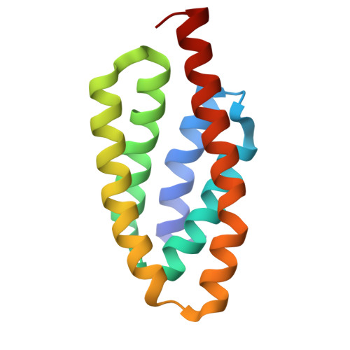
De novo enzyme design seeks to build proteins containing ideal active sites with catalytic residues surrounding and stabilizing the transition state(s) of the target chemical reaction 1-7 . The generative artificial intelligence method RFdiffusion 8,9 solves this problem, but requires specifying both the sequence position and backbone coordinates for each catalytic residue, limiting sampling. Here we introduce RFdiffusion2, which eliminates these requirements, and use it to design zinc metallohydrolases starting from quantum chemistry-derived active site geometries. From an initial set of 96 designs tested experimentally, the most active has a catalytic efficiency (k cat /K M ) of 16,000 M -1 s -1 , orders of magnitude higher than previously designed metallohydrolases 6,7,10,11 . A second round of 96 designs yielded 3 additional highly active enzymes, with k cat /K M values of up to 53,000 M -1 s -1 and a catalytic rate constant (k cat ) of up to 1.5 s -1 . The design models of the four most active designs differ from known structures and from each other, and the crystal structure of the most active design is very close to the design model, demonstrating the accuracy of the design method. The most active enzymes are predicted by PLACER 12 and Chai-1 (ref. 13 ) to have preorganized active sites that effectively position the substrate for nucleophilic attack by a water molecule activated by the bound metal. The ability to generate highly active enzymes directly from the computer, without experimental optimization, should enable a new generation of potent designer catalysts 14,15 .
- Department of Biochemistry, University of Washington, Seattle, WA, USA.
Organizational Affiliation: