In situ structural mechanism of epothilone-B-induced CNS axon regeneration.
Bodakuntla, S., Taira, K., Yamada, Y., Alvarez-Brecht, P., Cada, A.K., Basnet, N., Zhang, R., Martinez-Sanchez, A., Biertumpfel, C., Mizuno, N.(2025) Nature 648: 477-487
- PubMed: 41224993 Search on PubMedSearch on PubMed Central
- DOI: https://doi.org/10.1038/s41586-025-09654-z
- Primary Citation Related Structures:
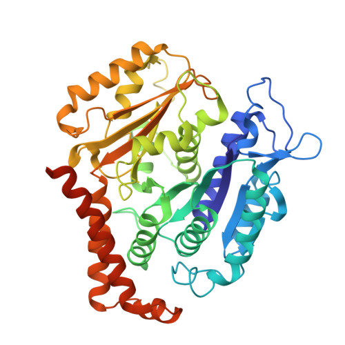
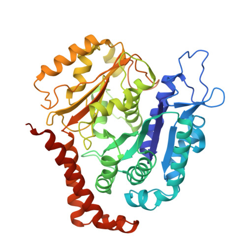
9PND - PubMed Abstract:
Axons in the adult central nervous system (CNS) do not regenerate following injury, in contrast to neurons in the peripheral nervous system and neuronal growth during embryonic development. The molecular mechanisms that prevent regeneration of neurons in the CNS remain largely unknown 1,2 . Here, to address the intracellular response to injury, we developed an in situ cryo-electron tomography and cryo-electron microscopy platform to mimic axonal damage and present the structural mechanism underlying thalamic axon regeneration induced by the drug epothilone B. We observed that stabilized microtubules extend beyond the injury site, generating membrane tension and driving membrane expansion. Cryo-electron microscopy reveals the in situ structure of microtubules at 3.19 Å resolution, which engage epothilone B within the microtubule lattice at the regenerating front. During repair, tubulin clusters are delivered and incorporated into polymerizing microtubules at the regenerating site. These microtubule shoots serve as scaffolds for various types of vesicles and endoplasmic reticulum, facilitating the supply of materials necessary for axon repair until membrane tension normalizes. We demonstrate the unexpected ability of neuronal cells to adjust to strain induced by epothilone B, which creates homeostatic imbalances and activates axons to regeneration mode.
- Laboratory of Structural Cell Biology, National Heart, Lung and Blood Institute, National Institutes of Health, Bethesda, MD, USA.
Organizational Affiliation: