Transgenic mouse-derived human monoclonal antibodies targeting EBV gp350 and gp42 provide basis for therapeutic development.
Chhan, C.B., Lang, K., Davis, A.R., Wan, Y.H., Aldridge, N.T., Kher, G., Scharffenberger, S.C., Hardy, S.R., Iureniev, R., Giltiay, N.V., Edwards, K.R., Radtke, S., Kiem, H.P., Pancera, M., McGuire, A.T.(2026) Cell Rep Med 7: 102618-102618
- PubMed: 41707657 Search on PubMedSearch on PubMed Central
- DOI: https://doi.org/10.1016/j.xcrm.2026.102618
- Primary Citation Related Structures:
9PF9 - PubMed Abstract:
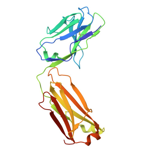
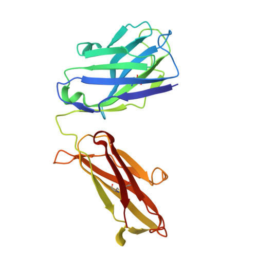
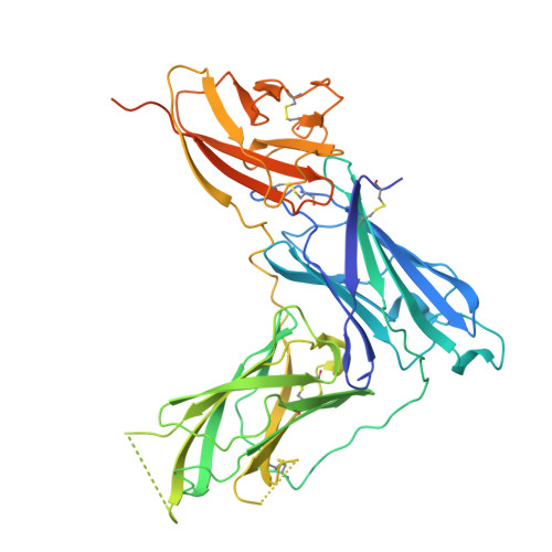
Epstein-Barr virus (EBV) causes infectious mononucleosis and contributes to neurodegenerative disorders and malignancies, particularly in immune-compromised hosts. Transplant patients face high risk of post-transplant lymphoproliferative disease, a life-threatening EBV-driven lymphoma. There are no EBV-specific vaccines or treatments; however, neutralizing antibodies against EBV glycoproteins may offer utility as therapeutic agents. EBV entry into B cells involves gp350, which binds complement receptors, and gp42, which engages HLA class II to trigger fusion. Most existing monoclonal antibodies (mAbs) against these antigens are non-human, limiting clinical use. Using a transgenic mouse model, we generate two gp350 and eight gp42 genetically human neutralizing mAbs that block receptor binding. Structural analyses reveal extended sites of vulnerability relevant to vaccine development. Delivery of a gp42 mAb protects humanized mice from EBV challenge, while a gp350 mAb provides partial protection. These mAbs highlight the utility of transgenic mice to produce therapeutic mAbs for preventing EBV-driven disease.
- Vaccine and Infectious Disease Division, Fred Hutchinson Cancer Center, Seattle, WA 98109, USA; Department of Global Health, University of Washington, Seattle, WA 98105, USA.
Organizational Affiliation: