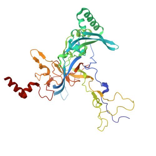
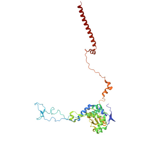
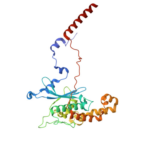
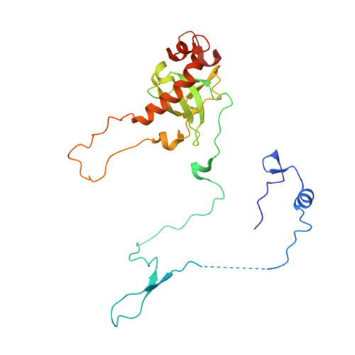
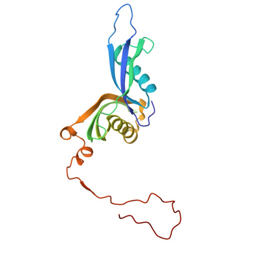
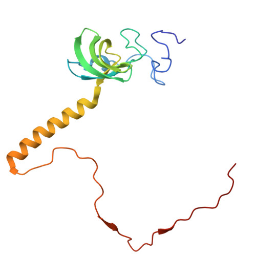
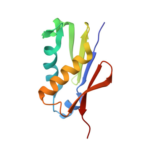
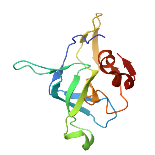
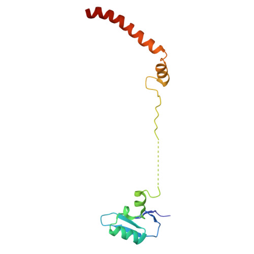
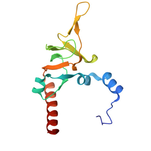
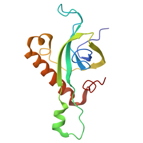
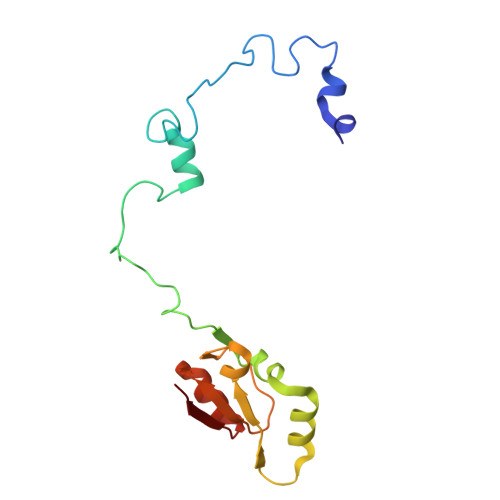
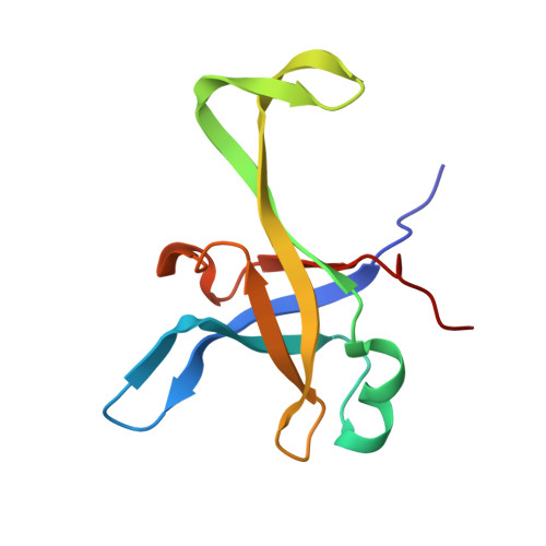
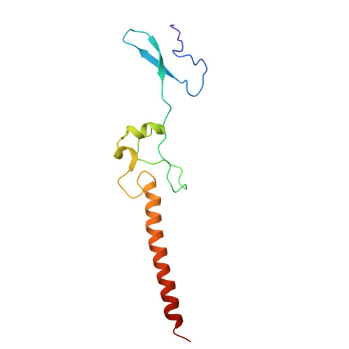
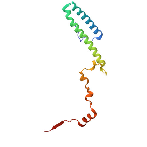
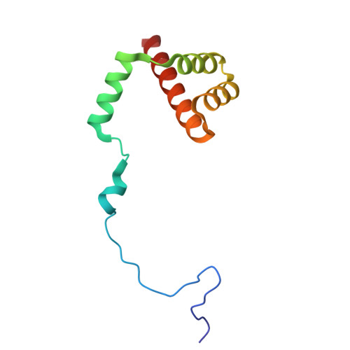
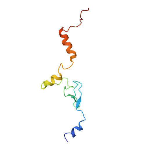
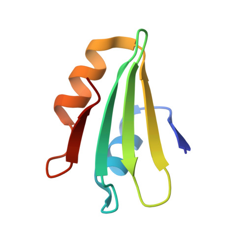
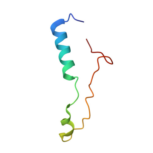
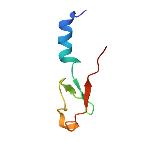
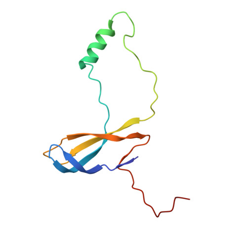
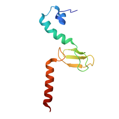
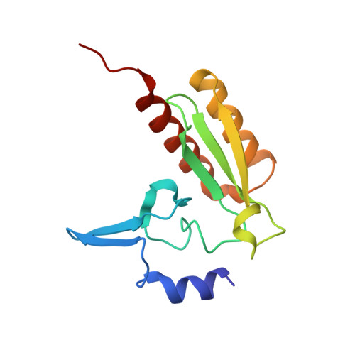
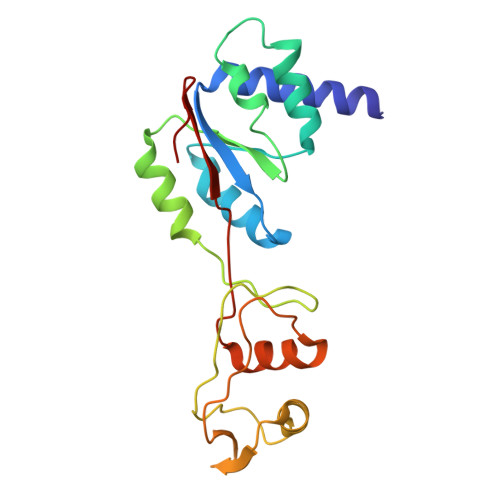
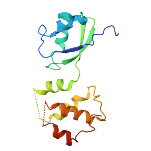
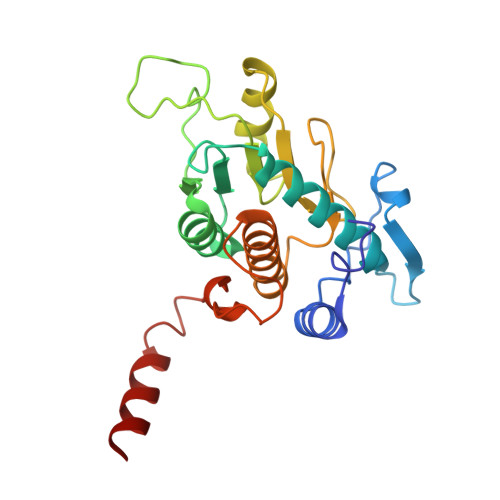
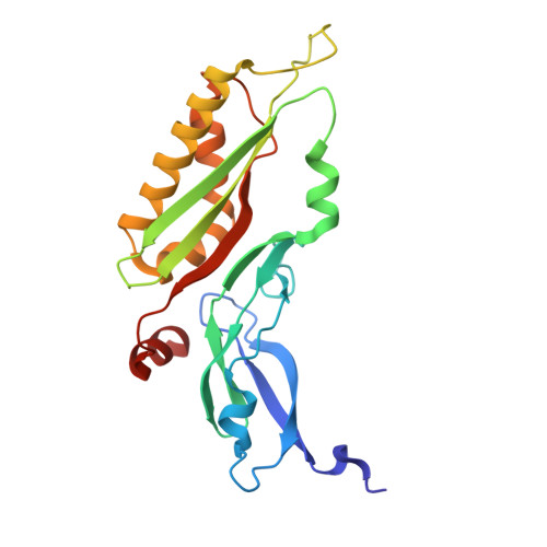
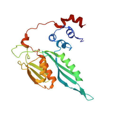
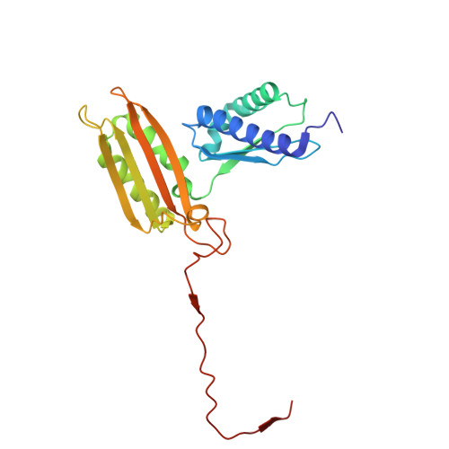
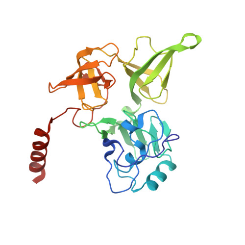
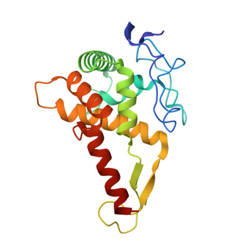
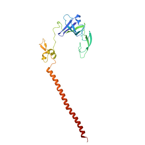
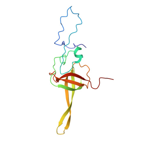
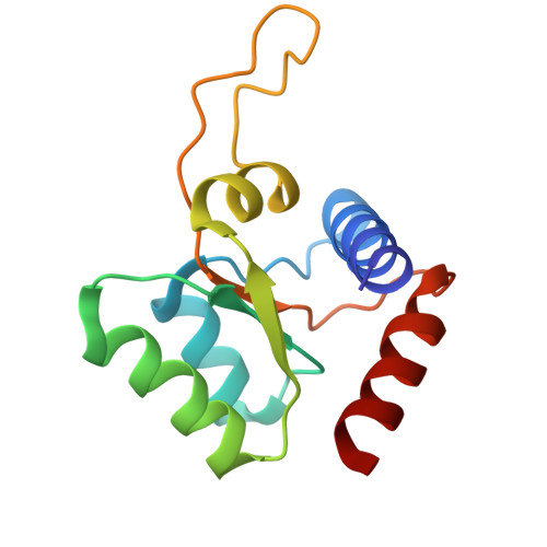
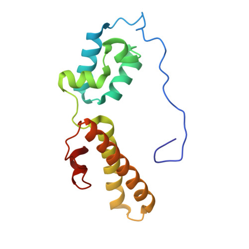
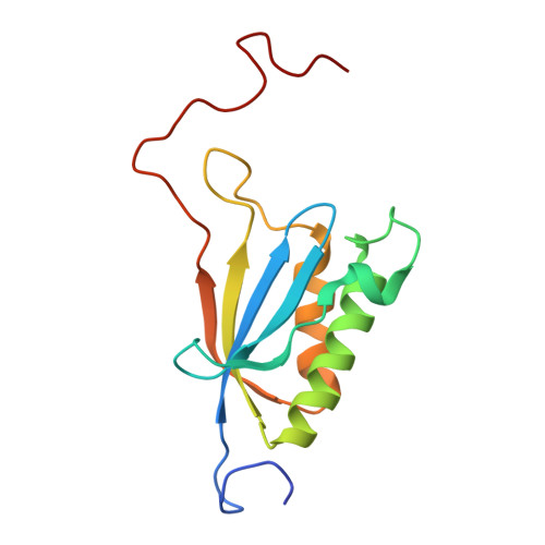
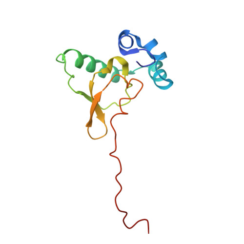
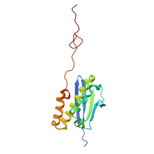
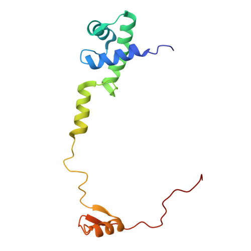
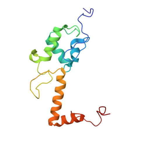
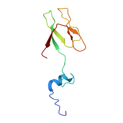
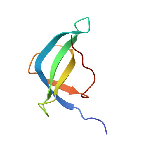
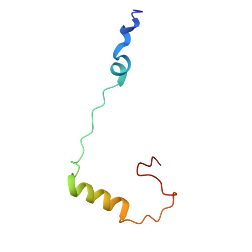
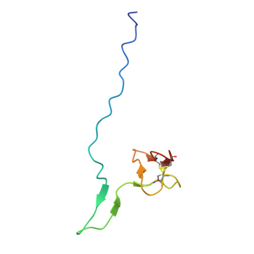
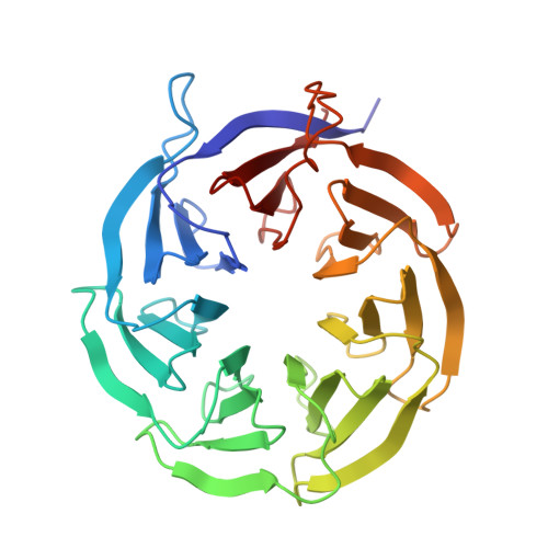
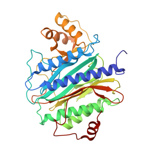
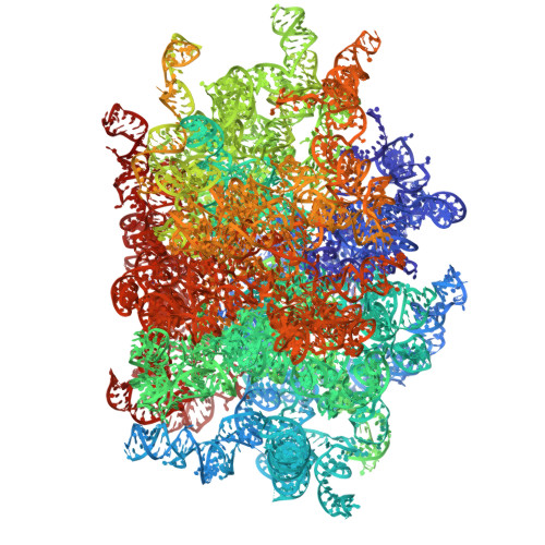
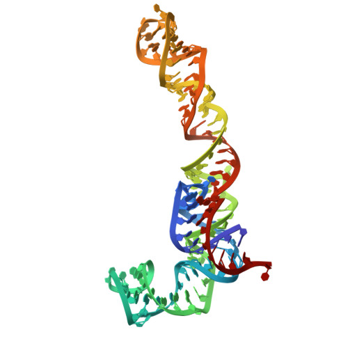
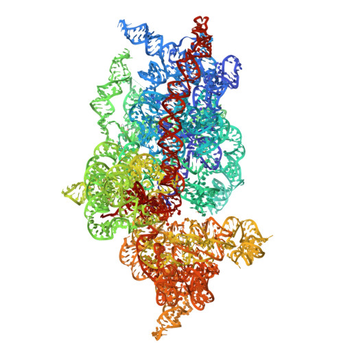
Comprehensive in situ structures of macromolecules can transform our understanding of biology and advance human health. Here, we map protein synthesis inside human cells in detail by combining automated cryo-focused ion beam (FIB) milling and in situ single-particle cryo electron microscopy (cryo-EM). With this in situ cryo-EM approach, we resolved a 2.2 Å consensus structure of the human 80S ribosome and unveiled 23 functional states, nearly all better than 3 Å resolution. Compared to in vitro studies, we observed variations in ribosome structures, distinct environments of ion and polyamine binding, and associated proteins such as EDF1 and NACβ that are typically not enriched with purified ribosomes. We also detected additional peptide-related density features on the ribosome and visualized ribosome-ribosome interactions in helical polysomes. Finally, high-resolution structures from cells treated with homoharringtonine and cycloheximide revealed a distinct translational landscape and a spermidine that interacts with cycloheximide at the E site, one of the numerous polyamines that also bind native ribosomes. These results underscore the value of high-resolution in situ studies in the native environment.
Organizational Affiliation:
Department of Molecular Biophysics and Biochemistry, Yale University, New Haven, CT, USA.
Microbial Sciences Institute, Yale University, West Haven, CT, USA.
Department of Microbial Pathogenesis, Yale University, New Haven, CT, USA.
Department of Pharmacology, Yale University, West Haven, CT, USA.
Department of Dermatology, Yale University, New Haven, CT, USA.
Department of Molecular Biophysics and Biochemistry, Yale University, New Haven, CT, USA. yong.xiong@yale.edu.