Conformational changes in ketohexokinase are conserved across isozymes and species.
Bae, S.Y., Allen, K.N., Tolan, D.R.(2025) Acta Crystallogr F Struct Biol Commun 81: 451-458
- PubMed: 41070915 Search on PubMedSearch on PubMed Central
- DOI: https://doi.org/10.1107/S2053230X25008428
- Primary Citation Related Structures:
9P6Q - PubMed Abstract:
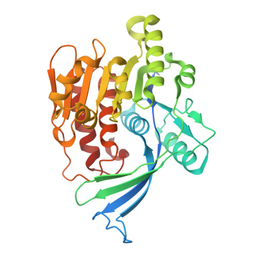
Ketohexokinase (KHK) catalyses the initial step in fructose metabolism, converting the furanose form of D-fructose to fructose 1-phosphate in an ATP-dependent reaction. Given its central role in metabolic pathways, KHK has emerged as a target for pharmacological intervention in the treatment of non-alcoholic fatty liver disease, metabolic syndrome, type 2 diabetes and obesity. KHK exists as two isoforms, A and C, which arise from alternative splicing of exon 3, resulting in a differing 45-amino-acid sequence within the 298-amino-acid primary structure of the enzyme. KHK is a biological homodimer, with each subunit adopting an α/β-fold architecture that interlocks with a β-clasp domain. In the case of KHK-C at least two distinct conformations of the β-clasp domain have been identified, whereas this conformational flexibility had not been observed in KHK-A. Here, X-ray crystallographic structural investigations of unliganded murine KHK-A refined to 1.37 Å resolution revealed the adoption of two conformations similar to those adopted by the human ortholog, suggesting that this structural feature is conserved across species. The functional significance of these conformational changes in KHK-A is of particular interest as this isoform has been implicated in cancer metastasis through a `moonlighting' protein kinase activity. Understanding the mechanistic role of conformational shifts in KHK-A may provide insights into its broader physiological functions and therapeutic potential.
- Program in Molecular Biology, Cell Biology, and Biochemistry, Boston University, Boston, MA 02215, USA.
Organizational Affiliation: