Download Mendeley
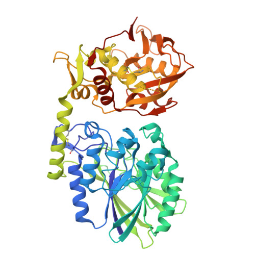
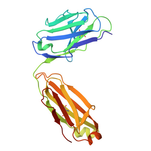
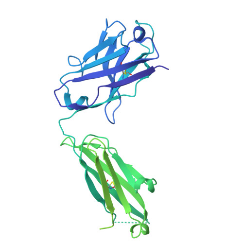
Sym024 Interacts with a Unique Epitope on the CD73 Homodimer, Favoring Effective Bivalent Binding to Improve Anti-PD-1 Therapy.
Jakobsen, J.S., Grandal, M.M., Hansen, R.W., Bansia, H., Armbruster, E., Theret, I., Skartved, N.J.O., Hald, R., Melander, M.C., Worsaae, A., Riva, M., Reckzeh, K., Vuillard, L., Lantto, J., des Georges, A., Frohlich, C.(2026) Clin Cancer Res 32: 1120-1135
- PubMed: 41511395 Search on PubMedSearch on PubMed Central
- DOI: https://doi.org/10.1158/1078-0432.CCR-25-2406
- Primary Citation Related Structures:
9P1M - PubMed Abstract:
Adenosine signaling may be a central immunosuppressive mechanism in several cancers, and blockade of the rate-limiting CD73 adenosine monophosphate (AMP)-to-adenosine enzyme has been demonstrated to improve the clinical efficacy of programmed cell death protein (ligand) 1 [PD-(L)1] immune therapy. However, deep inhibition of CD73 activity could prove difficult in tumor environments with a constant AMP supply and high CD73 levels. In this study, we sought to identify, characterize, and benchmark a novel antagonistic anti-CD73 antibody, Sym024 (S95024), and to structurally decode its mode of action. Sym024, selected via functional antibody repertoire screening, was tested against benchmark anti-CD73 antibodies in primary cell, cell line in vitro binding, CD73 enzymatic activity, and T-cell activation assays. Its in vivo tumor growth inhibition was examined in transplanted human or mouse tumors in immunocompetent or immunodeficient mice, and intratumoral enzymatic inhibition and immune cell recruitment were assessed. We investigated the Sym024-CD73 interaction using surface plasmon resonance, cryogenic electron microscopy, site-directed mutagenesis, and population-level complex formation through size-exclusion chromatography with light scatter mass detection. Preclinical safety and pharmacokinetics (PK) were assessed in monkeys. Sym024 effectively blocked CD73 across a large range of enzyme expression levels, comparing favorably with benchmark anti-CD73 antibodies; it improved the efficacy of PD-1 blockade in vitro as well as in vivo. Our structural data indicate that a unique one-to-one Sym024-CD73 interaction engenders this comprehensive inhibition. No preclinical safety flags were observed, and the PK profile of Sym024 supported a standard clinical dosing regimen. The comprehensive CD73 inhibition exhibited by Sym024 may improve the efficacy of anti-PD-(L)1/anti-CD73 combination treatment.
- Servier Symphogen A/S , Ballerup, Denmark.
Organizational Affiliation: