9ORH | pdb_00009orh

MicroED structure of the CTX-M-14 beta-lactamase-avibactam complex from inhibitor cocktail-soaked crystals


Experimental Data Snapshot

  • Method: ELECTRON CRYSTALLOGRAPHY
  • Resolution: 2.00 Å
  • R-Value Free: 
    0.245 (Depositor), 0.244 (DCC) 
  • R-Value Work: 
    0.197 (Depositor), 0.227 (DCC) 
  • R-Value Observed: 
    0.202 (Depositor) 

Starting Model: experimental
View more details

wwPDB Validation 3D Report Full Report

Validation slider image for 9ORH

This is version 1.1 of the entry. See complete history

Literature

Combining MicroED and native mass spectrometry for structural discovery of enzyme-small molecule complexes.

Vlahakis, N.W.Flowers, C.W.Liu, M.Agdanowski, M.P.Johnson, S.Summers, J.A.Jacobs, L.M.C.Keyser, C.Russell, P.Rose, S.L.Orlans, J.Adhami, N.Chen, Y.Sawaya, M.R.Basu, S.de Sanctis, D.Chen, Y.Wakatsuki, S.Nelson, H.M.Loo, J.A.Tang, Y.Rodriguez, J.A.

(2025) Proc Natl Acad Sci U S A 122: e2503780122-e2503780122

  • DOI: https://doi.org/10.1073/pnas.2503780122
  • Primary Citation Related Structures: 
    9NBP, 9NBQ, 9NC1, 9NCA, 9NCC, 9OQE, 9OR3, 9OR7, 9ORB, 9ORG, 9ORH, 9ORL, 9ORS, 9ORV, 9ORW, 9ORX, 9ORY, 9ORZ, 9OS0, 9OS1, 9OS8

  • PubMed Abstract: 

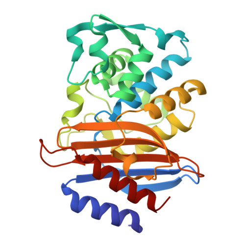
    With the goal of accelerating the discovery of small molecule-protein complexes, we leverage fast, low-dose, event-based electron counting microcrystal electron diffraction (MicroED) data collection and native mass spectrometry. This approach, which we term electron diffraction with native mass spectrometry (ED-MS), allows assignment of protein target structures bound to ligands with data obtained from crystal slurries soaked with mixtures of known inhibitors and crude biosynthetic reactions. This extends to libraries of printed ligands dispensed directly onto TEM grids for later soaking with microcrystal slurries, and complexes with noncovalent ligands. ED-MS resolves structures of the natural product, epoxide-based cysteine protease inhibitor E-64, and its biosynthetic analogs bound to the model cysteine protease, papain. It further identifies papain binding to its preferred natural products, by showing that two analogs of E-64 outcompete others in binding to papain crystals, and by detecting papain bound to E-64 and an analog from crude biosynthetic reactions, without purification. ED-MS also resolves binding of the CTX-M-14 β-lactamase, a target of active drug development, to the non-β-lactam inhibitor, avibactam, alone or in a cocktail of unrelated compounds. These results illustrate the utility of ED-MS for natural product ligand discovery and for structure-based screening of small molecule binders to macromolecular targets, promising utility for drug discovery.


  • Organizational Affiliation
    • Department of Chemistry and Biochemistry, University of California, Los Angeles, CA 90095.

Macromolecule Content 

  • Total Structure Weight: 56.28 kDa 
  • Atom Count: 4,022 
  • Modeled Residue Count: 524 
  • Deposited Residue Count: 524 
  • Unique protein chains: 1

Macromolecules

Find similar proteins by:|  3D Structure
Entity ID: 1
MoleculeChains  Sequence LengthOrganismDetailsImage
Beta-lactamase
A, B
262Escherichia coliMutation(s): 0 
Gene Names: 
EC: 3.5.2.6
UniProt
Find proteins for A0A649V088 (Escherichia coli)
Explore A0A649V088 
Go to UniProtKB:  A0A649V088
Entity Groups
Sequence Clusters30% Identity50% Identity70% Identity90% Identity95% Identity100% Identity
UniProt GroupA0A649V088
Sequence Annotations
Expand
Reference Sequence

Small Molecules

Ligands 1 Unique
IDChains Name / Formula / InChI Key2D Diagram3D Interactions
NXL

Query on NXL


Download:Ideal Coordinates CCD File
C [auth A],
D [auth B]
(2S,5R)-1-formyl-5-[(sulfooxy)amino]piperidine-2-carboxamide
C7 H13 N3 O6 S
WJDGWXPPFHLLNL-RITPCOANSA-N

Experimental Data & Validation

Experimental Data

  • Method: ELECTRON CRYSTALLOGRAPHY
  • Resolution: 2.00 Å
  • R-Value Free:  0.245 (Depositor), 0.244 (DCC) 
  • R-Value Work:  0.197 (Depositor), 0.227 (DCC) 
  • R-Value Observed: 0.202 (Depositor) 
Space Group: P 1 21 1
Unit Cell:
Length ( Å )Angle ( ˚ )
a = 44.9α = 90
b = 106.42β = 100.69
c = 47.52γ = 90
Software Package:
Software NamePurpose
PHENIXrefinement
XDSdata reduction
XSCALEdata scaling
PHASERphasing
PDB_EXTRACTdata extraction

Structure Validation

View Full Validation Report


Entry History 

& Funding Information

Deposition Data


Funding OrganizationLocationGrant Number
National Science Foundation (NSF, United States)United StatesDMR-1548924
Howard Hughes Medical Institute (HHMI)United StatesHHMI-EPI
Department of Energy (DOE, United States)United StatesDE-FC02-02ER63421

Revision History  (Full details and data files)

  • Version 1.0: 2025-06-18
    Type: Initial release
  • Version 1.1: 2025-08-13
    Changes: Database references