Funding Organization(s): National Institutes of Health/National Institute of General Medical Sciences (NIH/NIGMS), Japan Society for the Promotion of Science (JSPS)
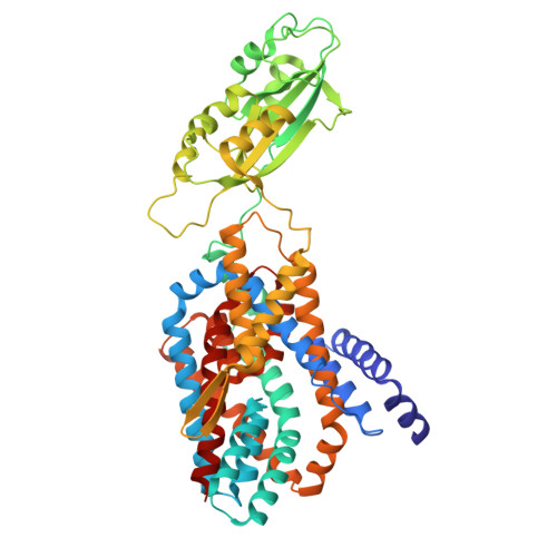
The transport of compounds across the cell membrane is essential for maintaining cellular homeostasis. Secondary exchange transporters mediate the movement of a wide range of substrates against their concentration gradients by harnessing the energy stored in electrochemical gradients. However, the molecular mechanism of substrate exchange by secondary transporters remains unclear. Here, we determined the structures of the aspartate exchanger AspT from Tetragenococcus halophilus using cryo-EM single-particle analysis and X-ray crystallography. We captured AspT in two distinct conformations: the apo outward-facing state and the substrate (L-Aspartate)-bound partially-open inward-facing intermediate state. AspT functions as a homodimer and comprises three domains: a dimerization domain, a substrate transport domain, and a soluble domain. Within each monomer, two hairpin loops in the transport domain form a single substrate-binding pocket. Upon L-aspartate binding, the transport domain carrying the substrate translocates toward the cytoplasmic side of the membrane, forming an outer barrier that blocks the periplasmic access to the binding pocket. These structural insights reveal that AspT mediates substrate translocation via an elevator-type alternating-access mechanism involving a stable partially-open inward-facing intermediate. By elucidating the mechanism of substrate exchange in secondary transporters, this study advances our understanding of membrane transport leading to translational applications in biotechnology.
Organizational Affiliation:
The Advanced Research Center for Innovations in Next-Generation Medicine (INGEM), Tohoku University, Sendai, Miyagi, Japan.
Tohoku Medical Megabank Organization, Tohoku University, Sendai, Miyagi, Japan.
Department of Microbial Resources, Graduate School of Agricultural Science, Tohoku University, Sendai, Miyagi, Japan.
Laboratory of Applied Microbiology, Department of Microbial Biotechnology, Graduate School of Agricultural Science, Tohoku University, Sendai, Miyagi, Japan.
Department of Cell Physiology and Molecular Biophysics, Center for Membrane Protein Research, School of Medicine, Texas Tech University Health Sciences Center, Lubbock, TX, USA.
Department of Cell Physiology and Molecular Biophysics, Center for Membrane Protein Research, School of Medicine, Texas Tech University Health Sciences Center, Lubbock, TX, USA. lan.guan@ttuhsc.edu.
Scientific Imaging Section, Research Support Division, Okinawa Institute of Science and Technology Graduate University (OIST), Onna-Son, Kunigami-Gun, Okinawa, Japan.
Quantum Wave Microscopy Unit, Okinawa Institute of Science and Technology Graduate University (OIST), Onna-Son, Kunigami-Gun, Okinawa, Japan.
Graduate School of Information Sciences, Tohoku University, Sendai, Miyagi, Japan.
Institute of Multidisciplinary Research for Advanced Materials, Tohoku University, Sendai, Miyagi, Japan.
Medical Institute of Bioregulation, Kyushu University, Fukuoka, Japan.
Biostructural Mechanism Laboratory, RIKEN SPring-8 Center, Sayo, Japan.
Department of Molecular Cell Science, Graduate School of Agricultural Science, Tohoku University, Sendai, Miyagi, Japan.
Department of Chemistry, Graduate School of Science, Chiba University, Chiba, Japan.
Membrane Protein Research Center, Chiba University, Chiba, Japan.
Institute for Advanced Academic Research, Chiba University, Chiba, Japan.
Research Center for Ultra-High Voltage Electron Microscopy, Osaka University, Osaka, Japan.
Department of Microbial Resources, Graduate School of Agricultural Science, Tohoku University, Sendai, Miyagi, Japan. keietsu.abe.b5@tohoku.ac.jp.
Laboratory of Applied Microbiology, Department of Microbial Biotechnology, Graduate School of Agricultural Science, Tohoku University, Sendai, Miyagi, Japan. keietsu.abe.b5@tohoku.ac.jp.
Microbial Genomics Laboratory, New Industry Creation Hatchery Center, Tohoku University, Sendai, Miyagi, Japan. keietsu.abe.b5@tohoku.ac.jp.
The Advanced Research Center for Innovations in Next-Generation Medicine (INGEM), Tohoku University, Sendai, Miyagi, Japan. masayuki.yamamoto.c7@tohoku.ac.jp.
Tohoku Medical Megabank Organization, Tohoku University, Sendai, Miyagi, Japan. masayuki.yamamoto.c7@tohoku.ac.jp.
The Advanced Research Center for Innovations in Next-Generation Medicine (INGEM), Tohoku University, Sendai, Miyagi, Japan. seizo.koshiba.b3@tohoku.ac.jp.
Tohoku Medical Megabank Organization, Tohoku University, Sendai, Miyagi, Japan. seizo.koshiba.b3@tohoku.ac.jp.