Delta-type glutamate receptors are ligand-gated ion channels.
Wang, H., Ahmed, F., Khau, J., Mondal, A.K., Twomey, E.C.(2025) Nature 647: 1063-1071
- PubMed: 40957579 Search on PubMed
- DOI: https://doi.org/10.1038/s41586-025-09610-x
- Primary Citation Related Structures:
9NWO, 9NWP, 9NWQ, 9OOO, 9OOP - PubMed Abstract:
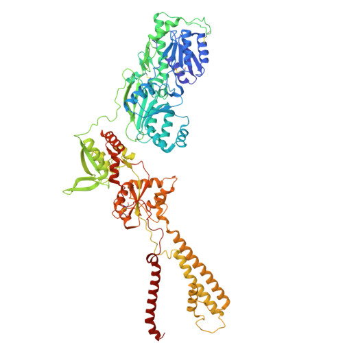
Delta-type ionotropic glutamate receptors (iGluRs), or GluDs, are members of the iGluR ligand-gated ion channel family, yet their function remains enigmatic 1 . Although GluDs are widely expressed in the brain, play key roles in synaptic organization, and harbor disease-linked mutations, whether they retain iGluR-like channel function is debated as currents have not been directly observed 2,3 . Here, we define GluDs as ligand-gated ion channels that are tightly regulated in cellular contexts by purifying human GluD2 (hGluD2) and directly characterizing its structure and function using cryo-electron microscopy (cryoEM) and bilayer recordings. We show that hGluD2 is activated by D-serine and γ-aminobutyric acid (GABA), with augmented activation at physiological temperatures. We reveal that hGluD2 contains an ion channel directly coupled to clamshell-like ligand-binding domains (LBDs), which are coordinated by the amino terminal domain (ATD) above the ion channel. Ligand binding triggers channel opening via an asymmetric mechanism, and a cerebellar ataxia point mutation in the LBD rearranges the receptor architecture and induces leak currents. Our findings demonstrate that GluDs possess the intrinsic biophysical properties of ligand-gated ion channels, reconciling prior conflicting observations to establish a framework for understanding their cellular regulation and for developing therapies targeting GluD2.
- Department of Biophysics and Biophysical Chemistry, Johns Hopkins University School of Medicine, Baltimore, MD, USA.
Organizational Affiliation: