9OIO | pdb_00009oio

The von Hippel Lindau-ElonginB-ElonginC (VCB) complex with fragments 9 and 14


Experimental Data Snapshot

  • Method: X-RAY DIFFRACTION
  • Resolution: 2.30 Å
  • R-Value Free: 
    0.249 (Depositor), 0.249 (DCC) 
  • R-Value Work: 
    0.211 (Depositor), 0.211 (DCC) 
  • R-Value Observed: 
    0.213 (Depositor) 

Starting Model: experimental
View more details

wwPDB Validation 3D Report Full Report

Validation slider image for 9OIO

Ligand Structure Quality Assessment 


This is version 1.1 of the entry. See complete history

Literature

NMR-Based Fragment Screen of the von Hippel-Lindau Elongin C&B Complex.

Amporndanai, K.Katinas, J.M.Chopra, A.Kayode, O.Vadukoot, A.K.Waterson, A.G.Fesik, S.W.

(2025) ACS Med Chem Lett 16: 1648-1654

  • DOI: https://doi.org/10.1021/acsmedchemlett.5c00316
  • Primary Citation Related Structures: 
    9OIM, 9OIN, 9OIO, 9OIQ

  • PubMed Abstract: 

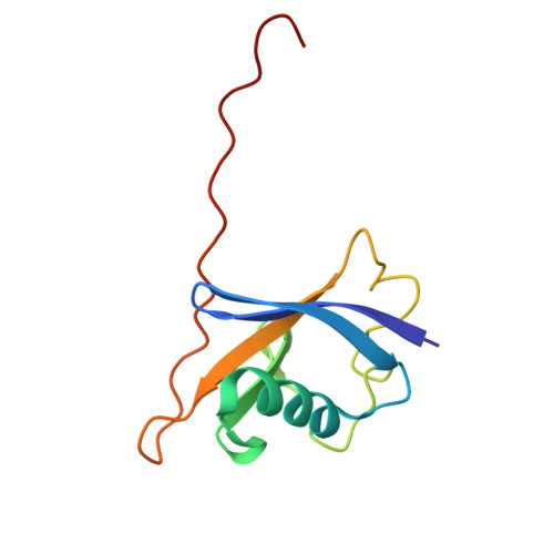
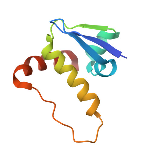
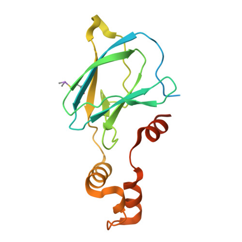
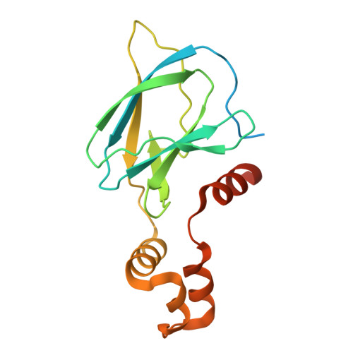
    von Hippel-Lindau (VHL) is an E3 ligase that has been widely exploited for the development of PROTACs to induce degradation of disease-associated target proteins. Nearly all VHL-recruiting PROTACs contain a hydroxyproline moiety based on the endogenous peptide substrate that occupies the HIF1α-binding site of VHL. However, the development of orally bioavailable PROTACs with hydroxyproline-based VHL ligands remains a significant hurdle, due to both the hydroxyproline and the peptidic nature of the VHL ligand. Here, we describe an NMR-based fragment screen against the VHL-Elongin C-Elongin B (VCB) complex. Several hits were shown by X-ray crystallography to bind to the HIF1α active site in VHL of the VCB complex, which opens the possibility for the discovery of new nonhydroxyproline-based VHL ligands for use in VHL-recruiting PROTACs.


  • Organizational Affiliation
    • Department of Biochemistry, Vanderbilt University School of Medicine, Nashville, Tennessee 37232-0146, United States.

Macromolecule Content 

  • Total Structure Weight: 176.15 kDa 
  • Atom Count: 10,766 
  • Modeled Residue Count: 1,322 
  • Deposited Residue Count: 1,528 
  • Unique protein chains: 4

Macromolecules

Find similar proteins by:|  3D Structure
Entity ID: 1
MoleculeChains  Sequence LengthOrganismDetailsImage
Elongin-B
A, D, G, J
104Homo sapiensMutation(s): 0 
Gene Names: ELOBTCEB2
UniProt & NIH Common Fund Data Resources
Find proteins for Q15370 (Homo sapiens)
Explore Q15370 
Go to UniProtKB:  Q15370
PHAROS:  Q15370
GTEx:  ENSG00000103363 
Entity Groups
Sequence Clusters30% Identity50% Identity70% Identity90% Identity95% Identity100% Identity
UniProt GroupQ15370
Sequence Annotations
Expand
Reference Sequence
Find similar proteins by:|  3D Structure
Entity ID: 2
MoleculeChains  Sequence LengthOrganismDetailsImage
Elongin-C
B, E, H, K
98Homo sapiensMutation(s): 0 
Gene Names: ELOCTCEB1
UniProt & NIH Common Fund Data Resources
Find proteins for Q15369 (Homo sapiens)
Explore Q15369 
Go to UniProtKB:  Q15369
PHAROS:  Q15369
GTEx:  ENSG00000154582 
Entity Groups
Sequence Clusters30% Identity50% Identity70% Identity90% Identity95% Identity100% Identity
UniProt GroupQ15369
Sequence Annotations
Expand
Reference Sequence
Find similar proteins by:|  3D Structure
Entity ID: 3
MoleculeChains  Sequence LengthOrganismDetailsImage
von Hippel-Lindau disease tumor suppressor
C, F, L
180Homo sapiensMutation(s): 0 
Gene Names: VHL
UniProt & NIH Common Fund Data Resources
Find proteins for P40337 (Homo sapiens)
Explore P40337 
Go to UniProtKB:  P40337
PHAROS:  P40337
GTEx:  ENSG00000134086 
Entity Groups
Sequence Clusters30% Identity50% Identity70% Identity90% Identity95% Identity100% Identity
UniProt GroupP40337
Sequence Annotations
Expand
Reference Sequence
Find similar proteins by:|  3D Structure
Entity ID: 4
MoleculeChains  Sequence LengthOrganismDetailsImage
von Hippel-Lindau disease tumor suppressor180Homo sapiensMutation(s): 0 
Gene Names: VHL
UniProt & NIH Common Fund Data Resources
Find proteins for P40337 (Homo sapiens)
Explore P40337 
Go to UniProtKB:  P40337
PHAROS:  P40337
GTEx:  ENSG00000134086 
Entity Groups
Sequence Clusters30% Identity50% Identity70% Identity90% Identity95% Identity100% Identity
UniProt GroupP40337
Sequence Annotations
Expand
Reference Sequence

Small Molecules

Ligands 3 Unique
IDChains Name / Formula / InChI Key2D Diagram3D Interactions
A1CBL
(Subject of Investigation/LOI)

Query on A1CBL


Download:Ideal Coordinates CCD File
M [auth C],
P [auth F],
W [auth L]
1-[(2-fluorophenyl)methyl]-4-(propan-2-yl)piperazine
C14 H21 F N2
SPOGRIJAQWZVBH-UHFFFAOYSA-N
A1CBK
(Subject of Investigation/LOI)

Query on A1CBK


Download:Ideal Coordinates CCD File
O [auth E],
S [auth H],
V [auth K]
1-[6-[oxidanyl(oxidanylidene)-$l^{4}-azanyl]-2,3-dihydroindol-1-yl]ethanone
C10 H10 N2 O3
RLXSSISTKOLICZ-UHFFFAOYSA-N
DMS

Query on DMS


Download:Ideal Coordinates CCD File
N [auth C]
Q [auth F]
R [auth F]
T [auth H]
U [auth I]
N [auth C],
Q [auth F],
R [auth F],
T [auth H],
U [auth I],
X [auth L]
DIMETHYL SULFOXIDE
C2 H6 O S
IAZDPXIOMUYVGZ-UHFFFAOYSA-N
Modified Residues  1 Unique
IDChains TypeFormula2D DiagramParent
CAS
Query on CAS
C, F, L
L-PEPTIDE LINKINGC5 H12 As N O2 SCYS

Experimental Data & Validation

Experimental Data

  • Method: X-RAY DIFFRACTION
  • Resolution: 2.30 Å
  • R-Value Free:  0.249 (Depositor), 0.249 (DCC) 
  • R-Value Work:  0.211 (Depositor), 0.211 (DCC) 
  • R-Value Observed: 0.213 (Depositor) 
Space Group: P 41 2 2
Unit Cell:
Length ( Å )Angle ( ˚ )
a = 92.904α = 90
b = 92.904β = 90
c = 359.384γ = 90
Software Package:
Software NamePurpose
PHENIXrefinement
XDSdata reduction
Aimlessdata scaling
PHASERphasing

Structure Validation

View Full Validation Report


Ligand Structure Quality Assessment 


Entry History 

& Funding Information

Deposition Data


Funding OrganizationLocationGrant Number
Other privateUnited StatesAWD00000788

Revision History  (Full details and data files)

  • Version 1.0: 2025-07-09
    Type: Initial release
  • Version 1.1: 2025-09-03
    Changes: Database references