Closed state structure of the pore revealed by uncoupled Shaker K + channel.
Liu, Y., Bassetto, C., Contreras, G.F., Perozo, E., Bezanilla, F.(2025) Nat Commun 16: 10180-10180
- PubMed: 41261088 Search on PubMedSearch on PubMed Central
- DOI: https://doi.org/10.1038/s41467-025-65124-0
- Primary Citation Related Structures:
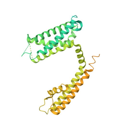
9OIC - PubMed Abstract:
Voltage gated potassium (Kv) channels regulate processes from cellular excitability to immune response and are major pharmaceutical targets. Despite recent structural advances, the closed state structure of the strictly coupled Kv1 family remains elusive. Here, we capture the structure of the Shaker potassium channel with a closed pore by uncoupling its voltage sensor domains from the pore domain. Structural determination of the uncoupled I384R mutant by single particle Cryo-EM reveals a fully closed pore coexisting with activated, non-relaxed voltage sensors. Comparison with the open pore structure suggests a roll-and-turn movement along the length of the pore-forming S6 helices, contrasting with canonical gating models based on limited movements of S6. These rotational-translational motions place two hydrophobic residues, one in the inner cavity and the other at the bundle crossing region, directly at the permeation pathway, limiting the pore radius to less than 1 Å. The selectivity filter is captured in a noncanonical state, partially expanded at G446, unlike previously described dilated or pinched filter conformations. Together, these findings suggest a reinterpretation of the mechanism of activation gating for strictly coupled Kv1 channels, highlighting the strictly sensor-pore coupling that underlies different functional states.
- Committee on Neurobiology, University of Chicago, Chicago, IL, USA.
Organizational Affiliation: