9OBP | pdb_00009obp

Cryo-EM structure of alpha-synuclein filaments derived from the frontal cortex of the case of atypical multiple system atrophy


Experimental Data Snapshot

  • Method: ELECTRON MICROSCOPY
  • Resolution: 2.50 Å
  • Aggregation State: FILAMENT 
  • Reconstruction Method: HELICAL 

wwPDB Validation   3D Report Full Report


This is version 1.1 of the entry. See complete history


Literature

Lewy-MSA hybrid fold drives distinct neuronal alpha-synuclein pathology.

Enomoto, M.Martinez-Valbuena, I.Forrest, S.L.Xu, X.Munhoz, R.P.Li, J.Rogaeva, E.Lang, A.E.Kovacs, G.G.

(2025) Commun Biol 8: 929-929

  • DOI: https://doi.org/10.1038/s42003-025-08355-7
  • Primary Citation of Related Structures:  
    9E9X, 9OBP

  • PubMed Abstract: 

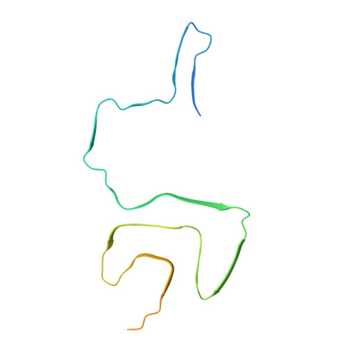
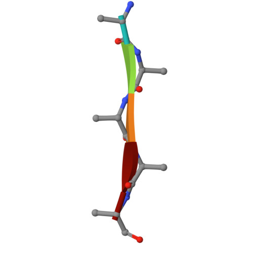
    The ordered assembly of α-synuclein protein encoded by SNCA into filaments characterizes neurodegenerative synucleinopathies. Lewy body disease (LBD) shows predominantly neuronal and multiple system atrophy (MSA), predominantly oligodendrocytic α-synuclein pathology affecting subcortical brain structures. Based on cryo-electron microscopy, it was reported that the structures of α-synuclein filaments from LBD differ from MSA and juvenile-onset synucleinopathy (JOS). The rare atypical MSA subtype shows abundant neuronal argyrophilic α-synuclein inclusions in the limbic system. Current concepts indicate that disease entities are characterized by unique protofilament folds. Here we demonstrate that α-synuclein can form a Lewy-MSA hybrid fold, leading to the atypical histopathological form of MSA. Distinct biochemical characteristics of α-synuclein, as demonstrated by protease-sensitivity digestion assay, seed amplification assays (SAAs), and conformational stability assays (CSA), are also linked to cytopathological differences. We expand the current structure-based classification of α-synucleinopathies and propose that cell-specific protein pathologies can be associated with distinct filament folds.


  • Organizational Affiliation
    • Princess Margaret Cancer Centre, University Health Network, Toronto, ON, Canada. Masahiro.Enomoto@uhn.ca.

Macromolecules
Find similar proteins by:  (by identity cutoff)  |  3D Structure
Entity ID: 1
MoleculeChains Sequence LengthOrganismDetailsImage
Alpha-synuclein
A, B, C, D, E
A, B, C, D, E, F, G, H, I, J
140Homo sapiensMutation(s): 0 
UniProt & NIH Common Fund Data Resources
Find proteins for P37840 (Homo sapiens)
Explore P37840 
Go to UniProtKB:  P37840
PHAROS:  P37840
GTEx:  ENSG00000145335 
Entity Groups  
Sequence Clusters30% Identity50% Identity70% Identity90% Identity95% Identity100% Identity
UniProt GroupP37840
Sequence Annotations
Expand
  • Reference Sequence

Find similar proteins by:  Sequence   |   3D Structure  

Entity ID: 2
MoleculeChains Sequence LengthOrganismDetailsImage
Unidentified protein
K, L, M, N, O
5Homo sapiensMutation(s): 0 
Sequence Annotations
Expand
  • Reference Sequence
Experimental Data & Validation

Experimental Data

  • Method: ELECTRON MICROSCOPY
  • Resolution: 2.50 Å
  • Aggregation State: FILAMENT 
  • Reconstruction Method: HELICAL 
EM Software:
TaskSoftware PackageVersion
MODEL REFINEMENTPHENIX1.21.2_5419

Structure Validation

View Full Validation Report


Entry History & Funding Information

Deposition Data


Funding OrganizationLocationGrant Number
National Institutes of Health/National Institute on Aging (NIH/NIA)United States--

Revision History  (Full details and data files)

  • Version 1.0: 2025-06-25
    Type: Initial release
  • Version 1.1: 2025-07-02
    Changes: Data collection, Database references