9O9U | pdb_00009o9u

The Erlin1/2 complex

  • Classification: MEMBRANE PROTEIN
  • Organism(s): Homo sapiens
  • Expression System: Homo sapiens
  • Mutation(s): No 

  • Deposited: 2025-04-18 Released: 2025-10-22 
  • Deposition Author(s): Gao, J., Shao, S.
  • Funding Organization(s): National Institutes of Health/National Institute on Aging (NIH/NIA), David and Lucile Packard Foundation, Richard and Susan Smith Family Foundation

Experimental Data Snapshot

  • Method: ELECTRON MICROSCOPY
  • Resolution: 3.00 Å
  • Aggregation State: PARTICLE 
  • Reconstruction Method: SINGLE PARTICLE 

wwPDB Validation   3D Report Full Report


This is version 1.1 of the entry. See complete history


Literature

Structures of human organellar SPFH protein complexes.

Gao, J.Sherpa, D.Kupko, N.Chino, H.Zeng, J.Shao, S.

(2025) Nat Commun 16: 10064-10064

  • DOI: https://doi.org/10.1038/s41467-025-65078-3
  • Primary Citation of Related Structures:  
    9O9U, 9O9Z, 9OA0

  • PubMed Abstract: 

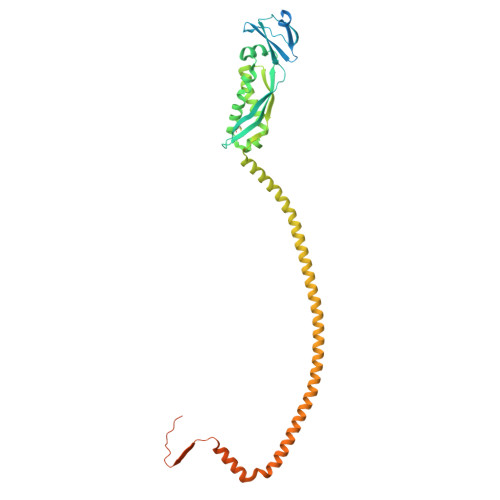
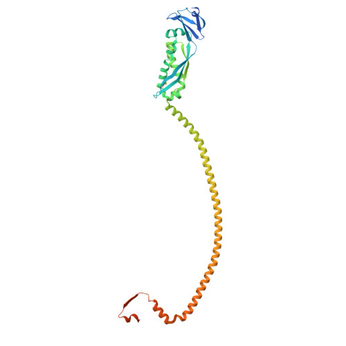
    Stomatin, Prohibitin, Flotillin, and HflK/C (SPFH) family proteins are found in all kingdoms of life and in multiple eukaryotic organelles. SPFH proteins assemble into homo- or hetero-oligomeric rings that form domed structures. Most SPFH assemblies also abut a cellular membrane, where they are implicated in diverse functions ranging from membrane organization to protein quality control. However, the precise architectures of different SPFH complexes remain unclear. Here, we report single-particle cryo-EM structures of the endoplasmic reticulum (ER)-resident Erlin1/2 complex and the mitochondrial prohibitin (PHB1/2) complex, revealing assemblies of 13 heterodimers of Erlin1 and Erlin2 and 11 heterodimers of PHB1 and PHB2, respectively. We also describe key interactions underlying the architecture of each complex and conformational heterogeneity of the PHB1/2 complex. Our findings elucidate the distinct stoichiometries and properties of human organellar SPFH complexes and highlight common principles of SPFH complex organization.


  • Organizational Affiliation
    • Department of Cell Biology, Harvard Medical School, Boston, MA, US.

Macromolecules
Find similar proteins by:  (by identity cutoff)  |  3D Structure
Entity ID: 1
MoleculeChains Sequence LengthOrganismDetailsImage
Erlin-1381Homo sapiensMutation(s): 0 
Gene Names: ERLIN1C10orf69KE04KEO4SPFH1
UniProt & NIH Common Fund Data Resources
Find proteins for O75477 (Homo sapiens)
Explore O75477 
Go to UniProtKB:  O75477
PHAROS:  O75477
GTEx:  ENSG00000107566 
Entity Groups  
Sequence Clusters30% Identity50% Identity70% Identity90% Identity95% Identity100% Identity
UniProt GroupO75477
Glycosylation
Glycosylation Sites: 1Go to GlyGen: O75477-1
Sequence Annotations
Expand
  • Reference Sequence
Find similar proteins by:  (by identity cutoff)  |  3D Structure
Entity ID: 2
MoleculeChains Sequence LengthOrganismDetailsImage
Erlin-2339Homo sapiensMutation(s): 0 
Gene Names: ERLIN2C8orf2SPFH2UNQ2441/PRO5003/PRO9924
UniProt & NIH Common Fund Data Resources
Find proteins for O94905 (Homo sapiens)
Explore O94905 
Go to UniProtKB:  O94905
PHAROS:  O94905
GTEx:  ENSG00000147475 
Entity Groups  
Sequence Clusters30% Identity50% Identity70% Identity90% Identity95% Identity100% Identity
UniProt GroupO94905
Glycosylation
Glycosylation Sites: 1Go to GlyGen: O94905-1
Sequence Annotations
Expand
  • Reference Sequence
Small Molecules
Ligands 1 Unique
IDChains Name / Formula / InChI Key2D Diagram3D Interactions
NAG (Subject of Investigation/LOI)
Query on NAG

Download Ideal Coordinates CCD File 
AA [auth A]
BA [auth a]
CA [auth B]
DA [auth b]
EA [auth C]
AA [auth A],
BA [auth a],
CA [auth B],
DA [auth b],
EA [auth C],
FA [auth c],
GA [auth D],
HA [auth d],
IA [auth E],
JA [auth e],
KA [auth F],
LA [auth f],
MA [auth G],
NA [auth g],
OA [auth H],
PA [auth h],
QA [auth I],
RA [auth i],
SA [auth J],
TA [auth j],
UA [auth K],
VA [auth k],
WA [auth L],
XA [auth l],
YA [auth M],
ZA [auth m]
2-acetamido-2-deoxy-beta-D-glucopyranose
C8 H15 N O6
OVRNDRQMDRJTHS-FMDGEEDCSA-N
Experimental Data & Validation

Experimental Data

  • Method: ELECTRON MICROSCOPY
  • Resolution: 3.00 Å
  • Aggregation State: PARTICLE 
  • Reconstruction Method: SINGLE PARTICLE 

Structure Validation

View Full Validation Report


Entry History & Funding Information

Deposition Data


Funding OrganizationLocationGrant Number
National Institutes of Health/National Institute on Aging (NIH/NIA)United StatesAG073277
David and Lucile Packard FoundationUnited States--
Richard and Susan Smith Family FoundationUnited States--

Revision History  (Full details and data files)

  • Version 1.0: 2025-10-22
    Type: Initial release
  • Version 1.1: 2025-12-24
    Changes: Data collection, Database references