Covalent Drug Binding in Live Cells Monitored by Mid-IR Quantum Cascade Laser Spectroscopy: Photoactive Yellow Protein as a Model System.
Mukherjee, S., Fried, S.D.E., Hong, N.Y., Bagheri, N., Kozuch, J., Mathews, I.I., Kirsh, J.M., Boxer, S.G.(2025) bioRxiv
- PubMed: 40894761 Search on PubMedSearch on PubMed Central
- DOI: https://doi.org/10.1101/2025.08.15.670201
- Primary Citation Related Structures:
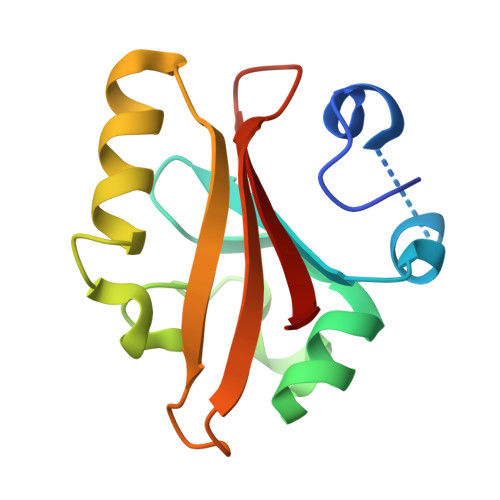
9O8V - PubMed Abstract:
The detection of drug-target interactions in live cells enables analysis of therapeutic compounds in a native cellular environment. Recent advances in spectroscopy and molecular biology have facilitated the development of genetically encoded vibrational probes like nitriles that can sensitively report on molecular interactions. Nitriles are powerful tools for measuring electrostatic environments within condensed media like proteins, but such measurements in live cells have been hindered by low signal-to-noise ratios. In this study, we design a spectrometer based on a double-beam quantum cascade laser (QCL)-based transmission infrared (IR) source with balanced detection that can significantly enhance sensitivity to nitrile vibrational probes embedded in proteins within cells compared to a conventional FTIR spectrometer. Using this approach, we detect small-molecule binding in E. coli , with particular focus on the interaction between para-coumaric acid (pCA) and nitrile-incorporated photoactive yellow protein (PYP). This system effectively serves as a model for investigating covalent drug binding in a cellular environment. Notably, we observe large spectral shifts of up to 15 cm -1 for nitriles embedded in PYP between the unbound and drug-bound states directly within bacteria, in agreement with observations for purified proteins. Such large spectral shifts are ascribed to the changes in the hydrogen-bonding environment around the local environment of nitriles, accurately modeled through high-level molecular dynamics simulations using the AMOEBA force field. Our findings underscore the QCL spectrometer's ability to enhance sensitivity for monitoring drug-protein interactions, offering new opportunities for advanced methodologies in drug development and biochemical research.
- Department of Chemistry, Stanford University, Stanford CA 94305, USA.
Organizational Affiliation: