9NXE | pdb_00009nxe

Crystal Structure of Calcineurin Clinical Variant E282K


Experimental Data Snapshot

  • Method: X-RAY DIFFRACTION
  • Resolution: 2.09 Å
  • R-Value Free: 
    0.243 (Depositor), 0.244 (DCC) 
  • R-Value Work: 
    0.202 (Depositor), 0.202 (DCC) 
  • R-Value Observed: 
    0.204 (Depositor) 

Starting Model: experimental
View more details

wwPDB Validation 3D Report Full Report

Validation slider image for 9NXE

This is version 1.1 of the entry. See complete history

Literature

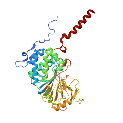
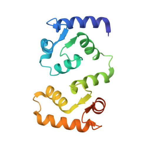
The clinical missense variant E282K in PPP3CA/calcineurin shifts substrate dephosphorylation by altering active site recruitment.

Shirakawa, K.T.Parikh, T.Machado, L.E.S.F.Poimenidou, G.Nguyen, H.T.Dell'Acqua, M.L.Kettenbach, A.N.Page, R.Peti, W.

(2026) Nat Commun 17

  • DOI: https://doi.org/10.1038/s41467-026-69535-5
  • Primary Citation Related Structures: 
    9NXE, 9NXF, 9NXN

  • PubMed Abstract: 

    Recently, de novo heterozygous variants of Calcineurin (CN) were reported as the cause of a neurodevelopmental disorder that presents with epileptic encephalopathy and dysmorphism (DEE91), with the largest group of patients harboring the CN missense mutation E282K (glutamate → lysine). Here, we use molecular and cellular techniques to define how this mutation alters CN activity. We discover that basophilic substrates use an arginine residue to bind to CN via an acidic substrate recruitment pocket adjacent to the CN active site, the E282 pocket. Furthermore, we show that basic residues in the i-1 position of the substrate relative to the substrate phosphosite enhance CN-mediated dephosphorylation. While the CN E282K structure shows that the overall conformation is unchanged, the E282 pocket transforms from acidic to basic, with pocket access blocked by the formation of a E282K-E237 salt bridge. Finally, in vitro assays and in cell phosphoproteomics show that CN E282K shifts CN substrate dephosphorylation profiles from basic to acidic, thereby altering CN-mediated dephosphorylation signaling. Together, these data define the molecular impact of the CN E282K variant in cells and development, providing a key step for developing strategies to treat this disorder and its accompanying complications.


  • Organizational Affiliation
    • Department of Molecular Biology and Biophysics, University of Connecticut Health Center, Farmington, CT, USA.

Macromolecule Content 

  • Total Structure Weight: 66.07 kDa 
  • Atom Count: 4,470 
  • Modeled Residue Count: 524 
  • Deposited Residue Count: 574 
  • Unique protein chains: 3

Macromolecules

Find similar proteins by:|  3D Structure
Entity ID: 1
MoleculeChains  Sequence LengthOrganismDetailsImage
Protein phosphatase 3 catalytic subunit alpha370Homo sapiensMutation(s): 1 
Gene Names: PPP3CACALNACNA
EC: 3.1.3.16
UniProt & NIH Common Fund Data Resources
Find proteins for Q08209 (Homo sapiens)
Explore Q08209 
Go to UniProtKB:  Q08209
PHAROS:  Q08209
GTEx:  ENSG00000138814 
Entity Groups
Sequence Clusters30% Identity50% Identity70% Identity90% Identity95% Identity100% Identity
UniProt GroupQ08209
Sequence Annotations
Expand
Reference Sequence
Find similar proteins by:|  3D Structure
Entity ID: 2
MoleculeChains  Sequence LengthOrganismDetailsImage
Calcineurin subunit B type 1156Homo sapiensMutation(s): 0 
Gene Names: PPP3R1CNA2CNB
UniProt & NIH Common Fund Data Resources
Find proteins for P63098 (Homo sapiens)
Explore P63098 
Go to UniProtKB:  P63098
PHAROS:  P63098
GTEx:  ENSG00000221823 
Entity Groups
Sequence Clusters30% Identity50% Identity70% Identity90% Identity95% Identity100% Identity
UniProt GroupP63098
Sequence Annotations
Expand
Reference Sequence
Find similar proteins by:|  3D Structure
Entity ID: 3
MoleculeChains  Sequence LengthOrganismDetailsImage
Sodium/hydrogen exchanger 148Homo sapiensMutation(s): 0 
Gene Names: SLC9A1APNH1NHE1
UniProt & NIH Common Fund Data Resources
Find proteins for P19634 (Homo sapiens)
Explore P19634 
Go to UniProtKB:  P19634
PHAROS:  P19634
GTEx:  ENSG00000090020 
Entity Groups
Sequence Clusters30% Identity50% Identity70% Identity90% Identity95% Identity100% Identity
UniProt GroupP19634
Sequence Annotations
Expand
Reference Sequence

Small Molecules

Ligands 4 Unique
IDChains Name / Formula / InChI Key2D Diagram3D Interactions
PEG

Query on PEG


Download:Ideal Coordinates CCD File
D [auth A]DI(HYDROXYETHYL)ETHER
C4 H10 O3
MTHSVFCYNBDYFN-UHFFFAOYSA-N
ZN

Query on ZN


Download:Ideal Coordinates CCD File
F [auth A]ZINC ION
Zn
PTFCDOFLOPIGGS-UHFFFAOYSA-N
FE

Query on FE


Download:Ideal Coordinates CCD File
E [auth A]FE (III) ION
Fe
VTLYFUHAOXGGBS-UHFFFAOYSA-N
CA

Query on CA


Download:Ideal Coordinates CCD File
G [auth B],
H [auth B],
I [auth B],
J [auth B]
CALCIUM ION
Ca
BHPQYMZQTOCNFJ-UHFFFAOYSA-N

Experimental Data & Validation

Experimental Data

  • Method: X-RAY DIFFRACTION
  • Resolution: 2.09 Å
  • R-Value Free:  0.243 (Depositor), 0.244 (DCC) 
  • R-Value Work:  0.202 (Depositor), 0.202 (DCC) 
  • R-Value Observed: 0.204 (Depositor) 
Space Group: C 2 2 21
Unit Cell:
Length ( Å )Angle ( ˚ )
a = 79.49α = 90
b = 126.501β = 90
c = 127.657γ = 90
Software Package:
Software NamePurpose
PHENIXrefinement
autoPROCdata reduction
autoPROCdata scaling
PHASERphasing

Structure Validation

View Full Validation Report


Entry History 

& Funding Information

Deposition Data


Funding OrganizationLocationGrant Number
National Institutes of Health/National Institute of General Medical Sciences (NIH/NIGMS)United StatesR01GM144379
National Institutes of Health/National Institute of General Medical Sciences (NIH/NIGMS)United StatesR01NS124666

Revision History  (Full details and data files)

  • Version 1.0: 2025-12-17
    Type: Initial release
  • Version 1.1: 2026-07-01
    Changes: Database references