9NWU | pdb_00009nwu

Crystal structure of a high affinity VL-VH tetrabody for the erythropoietin receptor


Experimental Data Snapshot

  • Method: X-RAY DIFFRACTION
  • Resolution: 1.96 Å
  • R-Value Free: 
    0.215 (Depositor), 0.216 (DCC) 
  • R-Value Work: 
    0.167 (Depositor), 0.167 (DCC) 
  • R-Value Observed: 
    0.169 (Depositor) 

Starting Model: experimental
View more details

wwPDB Validation 3D Report Full Report

Validation slider image for 9NWU

This is version 1.0 of the entry. See complete history

Literature

An asymmetric tetrabody is a potent and efficacious agonist of the erythropoietin receptor in vitro and in vivo.

Adams, J.J.Blazer, L.L.Chung, J.Karimi, M.Davidson, T.Bruce, H.A.Singer, A.U.Yang, N.Cardarelli, L.Pot, I.Colombo, L.Huang, L.J.Ma, Y.Michnick, S.W.Moe, O.W.Sidhu, S.S.

(2025) Protein Sci 34: e70292-e70292

  • DOI: https://doi.org/10.1002/pro.70292
  • Primary Citation Related Structures: 
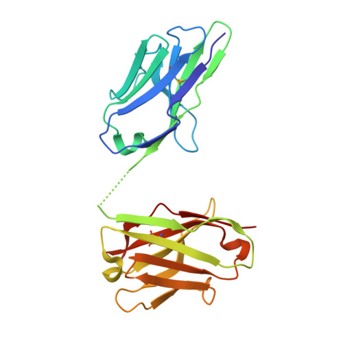
    9NWU

  • PubMed Abstract: 

    Erythropoietin (EPO) initiates EPO receptor (EPOR) signaling in hematopoietic cells by binding to an asymmetric EPOR dimer through two different sites. We engineered dimeric diabody-Fc (Db-Fc) fusion proteins that appeared to act as potent agonists of human EPOR in cell proliferation assays. However, detailed analysis of their oligomeric forms revealed that the predominant Db-Fc species bound EPOR with high affinity but failed to induce cell proliferation. Instead, a minor oligomeric form, identified as a putative tetrabody (Tb) fused to two Fc domains (Tb-Fc 2 ), proved to be the minimal active form. The existence of a tetrameric agonist was further supported by crystallography, which revealed an asymmetric Tb structure. Additionally, the structure of an antigen-binding fragment (Fab) bound to EPOR revealed an epitope distinct from the EPO binding sites, and structural modeling showed that engagement of two of the four binding sites on the Tb could form an asymmetric EPOR dimer nearly identical to the active conformation recruited by EPO. In a knock-in mouse model, where mouse EPOR was replaced by human EPOR, purified Tb-Fc 2 stimulated erythropoiesis with greater potency, efficacy, and duration than darbepoetin, a recombinant EPO that is the leading therapeutic erythropoiesis-stimulating agent (ESA). Collectively, these findings demonstrate that asymmetric tetravalent antibodies such as Tb-Fc 2 represent promising next-generation ESAs that provide enhanced potency, efficacy, and durability. Moreover, they may reduce the oncogenic and cardiovascular risks associated with the pleiotropy of EPO.


  • Organizational Affiliation
    • Anvil Institute for Systems Biologics, Canada.

Macromolecule Content 

  • Total Structure Weight: 99.9 kDa 
  • Atom Count: 7,580 
  • Modeled Residue Count: 903 
  • Deposited Residue Count: 916 
  • Unique protein chains: 1

Macromolecules

Find similar proteins by:|  3D Structure
Entity ID: 1
MoleculeChains  Sequence LengthOrganismDetailsImage
DiabodyA [auth C],
B [auth D],
C [auth A],
D [auth B]
229Homo sapiensMutation(s): 0 

Small Molecules

Ligands 4 Unique
IDChains Name / Formula / InChI Key2D Diagram3D Interactions
NHE

Query on NHE


Download:Ideal Coordinates CCD File
E [auth C],
G [auth D],
J [auth A],
M [auth B]
2-[N-CYCLOHEXYLAMINO]ETHANE SULFONIC ACID
C8 H17 N O3 S
MKWKNSIESPFAQN-UHFFFAOYSA-N
SO4

Query on SO4


Download:Ideal Coordinates CCD File
H [auth D]SULFATE ION
O4 S
QAOWNCQODCNURD-UHFFFAOYSA-L
CL

Query on CL


Download:Ideal Coordinates CCD File
F [auth C]
K [auth A]
L [auth A]
N [auth B]
O [auth B]
F [auth C],
K [auth A],
L [auth A],
N [auth B],
O [auth B],
P [auth B],
Q [auth B]
CHLORIDE ION
Cl
VEXZGXHMUGYJMC-UHFFFAOYSA-M
NA

Query on NA


Download:Ideal Coordinates CCD File
I [auth D],
R [auth B]
SODIUM ION
Na
FKNQFGJONOIPTF-UHFFFAOYSA-N

Experimental Data & Validation

Experimental Data

  • Method: X-RAY DIFFRACTION
  • Resolution: 1.96 Å
  • R-Value Free:  0.215 (Depositor), 0.216 (DCC) 
  • R-Value Work:  0.167 (Depositor), 0.167 (DCC) 
  • R-Value Observed: 0.169 (Depositor) 
Space Group: P 1 21 1
Unit Cell:
Length ( Å )Angle ( ˚ )
a = 57.355α = 90
b = 141.779β = 90.76
c = 60.651γ = 90
Software Package:
Software NamePurpose
PHENIXrefinement
autoPROCdata reduction
Aimlessdata scaling
PHASERphasing

Structure Validation

View Full Validation Report


Entry History 

& Funding Information

Deposition Data


Funding OrganizationLocationGrant Number
Canadian Institutes of Health Research (CIHR)CanadaPJT-159640
Other privateBristol-Myers-Squibb

Revision History  (Full details and data files)

  • Version 1.0: 2025-10-01
    Type: Initial release