9NO0 | pdb_00009no0

hPNPase RNA loading state


Experimental Data Snapshot

  • Method: ELECTRON MICROSCOPY
  • Resolution: 2.08 Å
  • Aggregation State: PARTICLE 
  • Reconstruction Method: SINGLE PARTICLE 

Starting Model: in silico
View more details

wwPDB Validation   3D Report Full Report


This is version 1.1 of the entry. See complete history


Literature

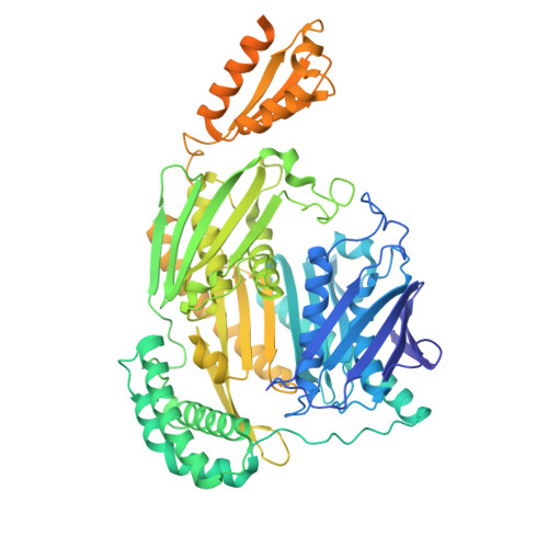
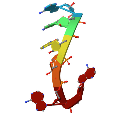
Loop-mediated regulation and base flipping drive RNA cleavage by human mitochondrial PNPase.

Unseld, O.Das, H.Hallberg, B.M.

(2025) Nucleic Acids Res 53

  • DOI: https://doi.org/10.1093/nar/gkaf1296
  • Primary Citation of Related Structures:  
    9NJB, 9NJC, 9NJD, 9NJE, 9NO0, 9XYI, 9XZF

  • PubMed Abstract: 

    Human polynucleotide phosphorylase (hPNPase), a trimeric exoribonuclease, is crucial for maintaining mitochondrial RNA metabolism, including the regulated degradation of RNA. Mutations in hPNPase have been linked to mitochondrial pathologies, underscoring its importance in mitochondrial RNA homeostasis. Despite this significance, the molecular basis of its catalytic mechanism and the structural consequences of active-site mutations remain poorly understood. We employed high-resolution electron cryo-microscopy to capture three distinct functional states of hPNPase during RNA degradation. In the loading state, flexible loops facilitate the recruitment of the substrate RNA and guide it toward the active site. During the pre-catalytic state, terminal nucleotides reorient within the active site, positioning the RNA backbone for cleavage, which is stabilized by Mg2+. Finally, the catalytic state reveals a nucleophilic attack of phosphate on the RNA backbone, mediated by key active-site residues. These results offer a clear biochemical framework for hPNPase-mediated RNA turnover, clarifying its catalytic mechanism and highlighting how active-site integrity is crucial for efficient RNA degradation.


  • Organizational Affiliation
    • Department of Cell and Molecular Biology, Karolinska Institutet, Stockholm  171 77, Sweden.

Macromolecules

Find similar proteins by:  (by identity cutoff)  |  3D Structure
Entity ID: 1
MoleculeChains Sequence LengthOrganismDetailsImage
Polyribonucleotide nucleotidyltransferase 1, mitochondrial
A, B, C
773Homo sapiensMutation(s): 0 
Gene Names: PNPT1PNPASE
EC: 2.7.7.8
UniProt & NIH Common Fund Data Resources
Find proteins for Q8TCS8 (Homo sapiens)
Explore Q8TCS8 
Go to UniProtKB:  Q8TCS8
PHAROS:  Q8TCS8
GTEx:  ENSG00000138035 
Entity Groups  
Sequence Clusters30% Identity50% Identity70% Identity90% Identity95% Identity100% Identity
UniProt GroupQ8TCS8
Sequence Annotations
Expand
  • Reference Sequence

Find similar nucleic acids by:  Sequence   |   3D Structure  

Entity ID: 2
MoleculeChains LengthOrganismImage
RNA analog6Homo sapiens
Sequence Annotations
Expand
  • Reference Sequence
Experimental Data & Validation

Experimental Data

  • Method: ELECTRON MICROSCOPY
  • Resolution: 2.08 Å
  • Aggregation State: PARTICLE 
  • Reconstruction Method: SINGLE PARTICLE 
EM Software:
TaskSoftware PackageVersion
RECONSTRUCTIONcryoSPARC4.6.2
MODEL REFINEMENTPHENIX1.21.2_5419

Structure Validation

View Full Validation Report


Entry History & Funding Information

Deposition Data


Funding OrganizationLocationGrant Number
Not funded--

Revision History  (Full details and data files)

  • Version 1.0: 2025-12-10
    Type: Initial release
  • Version 1.1: 2025-12-24
    Changes: Data collection, Database references