9NM3 | pdb_00009nm3

Crystal structure of FBF-1 RBD+CT complexed with compact FBE RNA


Experimental Data Snapshot

  • Method: X-RAY DIFFRACTION
  • Resolution: 2.65 Å
  • R-Value Free: 
    0.264 (Depositor), 0.264 (DCC) 
  • R-Value Work: 
    0.213 (Depositor), 0.213 (DCC) 
  • R-Value Observed: 
    0.218 (Depositor) 

Starting Model: experimental
View more details

wwPDB Validation   3D Report Full Report


This is version 1.1 of the entry. See complete history


Literature

Caenorhabditis elegans FBF-1 and FBF-2 C-terminal intrinsically disordered regions differentially regulate RNA-binding affinity.

Hawthorne, H.R.Qiu, C.Tanaka Hall, T.M.

(2025) RNA 31: 1391-1402

  • DOI: https://doi.org/10.1261/rna.080578.125
  • Primary Citation of Related Structures:  
    9NM3

  • PubMed Abstract: 

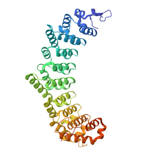
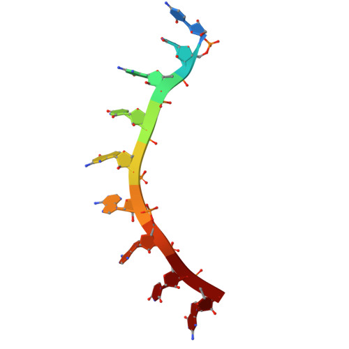
    PUF proteins (named for Drosophila melanogaster Pumilio and Caenorhabditis elegans fem-3 mRNA binding factor or FBF) are a family of RNA-binding proteins. C. elegans FBF is a collective term for two PUF proteins, FBF-1 and FBF-2, that maintain germline stem cells. FBF binds the 3'UTR of target RNAs and together with partner proteins represses translation of mRNAs that promote differentiation. Until recently, little was known about the functions of the FBF C-terminal intrinsically-disordered regions that follow the RNA-binding domain (RBD). Despite high overall protein sequence conservation (91% identical residues), the FBF-1 and FBF-2 C-terminal tails (CTs) are distinct, and the FBF-2 CT is essential for its function. The FBF-2 CT contains a PUF-interacting motif (PIM) that binds its own RBD and autoinhibits RNA-binding affinity. Here we investigated whether differences in the FBF-1 and FBF-2 CTs impact molecular function. Unlike FBF-2, the FBF-1 CT had no impact on RNA binding. Despite this, a crystal structure of FBF-1 demonstrated that a PIM in the FBF-1 CT binds to its RBD, like FBF-2. By creating FBF-1/FBF-2 chimeric proteins, we discovered that the FBF-2 CT can autoinhibit FBF-1 RNA binding, and substitution of the FBF-1 PIM for the FBF-2 PIM diminished FBF-2 autoinhibition. Our results exemplify how RBP paralogs diverge to fine tune their RNA-binding activities.


  • Organizational Affiliation
    • NIEHS, NIH.

Macromolecules

Find similar proteins by:  (by identity cutoff)  |  3D Structure
Entity ID: 1
MoleculeChains Sequence LengthOrganismDetailsImage
Fem-3 mRNA-binding factor 1454Caenorhabditis elegansMutation(s): 0 
Gene Names: fbf-1H12I13.4
UniProt
Find proteins for Q9N5M6 (Caenorhabditis elegans)
Explore Q9N5M6 
Go to UniProtKB:  Q9N5M6
Entity Groups  
Sequence Clusters30% Identity50% Identity70% Identity90% Identity95% Identity100% Identity
UniProt GroupQ9N5M6
Sequence Annotations
Expand
  • Reference Sequence

Find similar nucleic acids by:  Sequence   |   3D Structure  

Entity ID: 2
MoleculeChains LengthOrganismImage
RNA (5'-R(*CP*UP*GP*UP*GP*AP*AP*UP*G)-3')9Caenorhabditis elegans
Sequence Annotations
Expand
  • Reference Sequence
Experimental Data & Validation

Experimental Data

  • Method: X-RAY DIFFRACTION
  • Resolution: 2.65 Å
  • R-Value Free:  0.264 (Depositor), 0.264 (DCC) 
  • R-Value Work:  0.213 (Depositor), 0.213 (DCC) 
  • R-Value Observed: 0.218 (Depositor) 
Space Group: P 21 2 21
Unit Cell:
Length ( Å )Angle ( ˚ )
a = 42.832α = 90
b = 43.6β = 90
c = 253.278γ = 90
Software Package:
Software NamePurpose
PHENIXrefinement
HKL-2000data reduction
HKL-2000data scaling
PHASERphasing

Structure Validation

View Full Validation Report


Entry History & Funding Information

Deposition Data


Funding OrganizationLocationGrant Number
National Institutes of Health/National Institute of Environmental Health Sciences (NIH/NIEHS)United States--

Revision History  (Full details and data files)

  • Version 1.0: 2025-08-20
    Type: Initial release
  • Version 1.1: 2025-09-24
    Changes: Database references