9N65 | pdb_00009n65

Transporter associated with antigen processing (TAP) bound to ATP and ADP In the inward-facing conformation


Experimental Data Snapshot

  • Method: ELECTRON MICROSCOPY
  • Resolution: 3.70 Å
  • Resolution: 3.70 Å
  • Resolution: 3.70 Å
  • Resolution: 3.70 Å
  • Aggregation State: PARTICLE 
  • Reconstruction Method: SINGLE PARTICLE 

wwPDB Validation   3D Report Full Report


This is version 1.1 of the entry. See complete history


Literature

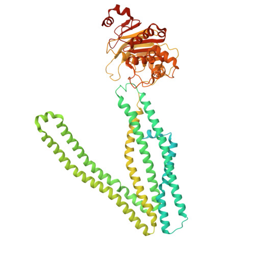
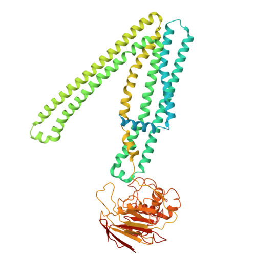
Nucleotide-dependent conformational changes direct peptide export by the transporter associated with antigen processing.

Lee, J.Manon, V.Chen, J.

(2025) Immunity 58: 2166-2175.e4

  • DOI: https://doi.org/10.1016/j.immuni.2025.08.003
  • Primary Citation Related Structures: 
    9N61, 9N62, 9N63, 9N64, 9N65, 9N66

  • PubMed Abstract: 

    The transporter associated with antigen processing (TAP) delivers peptide antigens from the cytoplasm into the endoplasmic reticulum (ER) for loading onto major histocompatibility complex class I (MHC-I) molecules. To examine the mechanisms of peptide transport and release into the ER, we determined cryo-electron microscopy structures of the human TAP heterodimer in multiple functional states along the transport cycle. In the inward-facing conformation, when the peptide translocation cavity within the TAP heterodimer is exposed to the cytosol, ATP binding strengthened intradomain assembly. Transition to the outward-facing conformation, when the transporter opens to the ER lumen, led to a complete reconfiguration of the peptide-binding site, facilitating peptide release. ATP hydrolysis opened the catalytically active nucleotide-binding consensus site, and the subsequent separation of the nucleotide-binding domains reset the transport cycle. These findings establish a comprehensive structural framework for understanding unilateral peptide transport, vanadate trapping, and trans-inhibition-an internal feedback mechanism that prevents excessive peptide accumulation and activation of the ER stress response.


  • Organizational Affiliation
    • Laboratory of Membrane Biophysics and Biology, the Rockefeller University, New York, NY 10065, USA.

Macromolecules
Find similar proteins by:  (by identity cutoff)  |  3D Structure
Entity ID: 1
MoleculeChains Sequence LengthOrganismDetailsImage
Antigen peptide transporter 1748Homo sapiensMutation(s): 0 
Gene Names: TAP1ABCB2PSF1RING4Y3
EC: 7.4.2.14
Membrane Entity: Yes 
UniProt & NIH Common Fund Data Resources
Find proteins for Q03518 (Homo sapiens)
Explore Q03518 
Go to UniProtKB:  Q03518
PHAROS:  Q03518
GTEx:  ENSG00000168394 
Entity Groups  
Sequence Clusters30% Identity50% Identity70% Identity90% Identity95% Identity100% Identity
UniProt GroupQ03518
Sequence Annotations
Expand
  • Reference Sequence
Find similar proteins by:  (by identity cutoff)  |  3D Structure
Entity ID: 2
MoleculeChains Sequence LengthOrganismDetailsImage
Antigen peptide transporter 2686Homo sapiensMutation(s): 0 
Gene Names: TAP2ABCB3PSF2RING11Y1
EC: 7.4.2.14
Membrane Entity: Yes 
UniProt & NIH Common Fund Data Resources
Find proteins for Q03519 (Homo sapiens)
Explore Q03519 
Go to UniProtKB:  Q03519
PHAROS:  Q03519
GTEx:  ENSG00000204267 
Entity Groups  
Sequence Clusters30% Identity50% Identity70% Identity90% Identity95% Identity100% Identity
UniProt GroupQ03519
Sequence Annotations
Expand
  • Reference Sequence
Experimental Data & Validation

Experimental Data

  • Method: ELECTRON MICROSCOPY
  • Resolution: 3.70 Å
  • Resolution: 3.70 Å
  • Resolution: 3.70 Å
  • Resolution: 3.70 Å
  • Aggregation State: PARTICLE 
  • Reconstruction Method: SINGLE PARTICLE 

Structure Validation

View Full Validation Report


Entry History & Funding Information

Deposition Data


Funding OrganizationLocationGrant Number
Howard Hughes Medical Institute (HHMI)United States--

Revision History  (Full details and data files)

  • Version 1.0: 2025-09-03
    Type: Initial release
  • Version 1.1: 2026-03-18
    Changes: Data collection, Database references