9MY3 | pdb_00009my3

Structure of Xenopus KCNQ1-CaM in GDN


Experimental Data Snapshot

  • Method: ELECTRON MICROSCOPY
  • Resolution: 3.46 Å
  • Aggregation State: PARTICLE 
  • Reconstruction Method: SINGLE PARTICLE 

wwPDB Validation   3D Report Full Report


This is version 1.0 of the entry. See complete history


Literature

A physiologically-relevant intermediate state structure of a voltage-gated potassium channel.

Kyriakis, E.Sastre, D.Eldstrom, J.Roscioni, A.Russo, S.Ataei, F.Dou, Y.Chan, M.Molinarolo, S.Maragliano, L.Van Petegem, F.Fedida, D.

(2025) Nat Commun 16: 8814-8814

  • DOI: https://doi.org/10.1038/s41467-025-64060-3
  • Primary Citation of Related Structures:  
    9MY3, 9MY4

  • PubMed Abstract: 

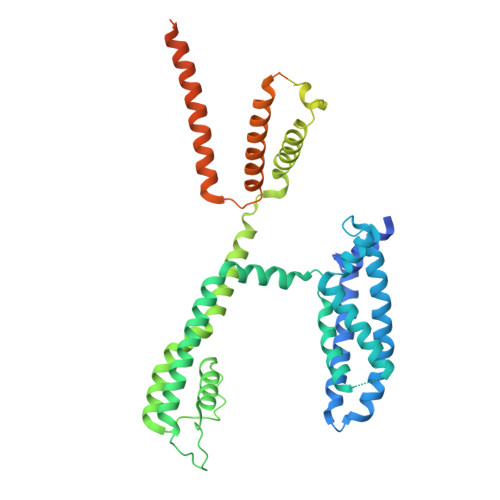
    Voltage-gated potassium ion (K + ) channels perform critical roles in many physiological processes, while gain- or loss-of-function mutations lead to life-threatening pathologies. Here, we establish the high-resolution structure of a pivotal intermediate state of the Kv7.1 (KCNQ1) channel using cryogenic electron microscopy. The 3.53 Å resolution structure reveals straightened upper S1 and S2 voltage sensor helices, distancing them from the pore filter helix compared to fully activated channels. The outward translation of the S4 voltage sensor is essentially complete in this intermediate state, and the S4-S6 helices and the S4-S5 linker do not change position significantly between intermediate and activated states. The PIP2 ligand can bind in both states. Movement of S1 and S2 helices towards the filter helix from intermediate to activated states may explain smaller components of KCNQ1 voltage sensor fluorescence, differential Rb + /K + selectivity, and pharmacological responses to activators and inhibitors. Single channel recordings and the location of long QT mutations suggest the potential physiological and disease importance of the intermediate state.


  • Organizational Affiliation
    • The Department of Anesthesiology, Pharmacology and Therapeutics, University of British Columbia, Vancouver, V6T 1Z3, Canada.

Macromolecules
Find similar proteins by:  (by identity cutoff)  |  3D Structure
Entity ID: 1
MoleculeChains Sequence LengthOrganismDetailsImage
Potassium voltage-gated channel subfamily KQT member 1
A, C, E, G
545Xenopus laevisMutation(s): 0 
Gene Names: kcnq1kvlqt1
Membrane Entity: Yes 
UniProt
Find proteins for P70057 (Xenopus laevis)
Explore P70057 
Go to UniProtKB:  P70057
Entity Groups  
Sequence Clusters30% Identity50% Identity70% Identity90% Identity95% Identity100% Identity
UniProt GroupP70057
Sequence Annotations
Expand
  • Reference Sequence
Find similar proteins by:  (by identity cutoff)  |  3D Structure
Entity ID: 2
MoleculeChains Sequence LengthOrganismDetailsImage
Calmodulin-1
B, D, F, H
149Homo sapiensMutation(s): 0 
Gene Names: CALM1CALMCAMCAM1
UniProt & NIH Common Fund Data Resources
Find proteins for P0DP23 (Homo sapiens)
Explore P0DP23 
Go to UniProtKB:  P0DP23
PHAROS:  P0DP23
GTEx:  ENSG00000198668 
Entity Groups  
Sequence Clusters30% Identity50% Identity70% Identity90% Identity95% Identity100% Identity
UniProt GroupP0DP23
Sequence Annotations
Expand
  • Reference Sequence
Experimental Data & Validation

Experimental Data

  • Method: ELECTRON MICROSCOPY
  • Resolution: 3.46 Å
  • Aggregation State: PARTICLE 
  • Reconstruction Method: SINGLE PARTICLE 
EM Software:
TaskSoftware PackageVersion
RECONSTRUCTIONcryoSPARC4.2.1

Structure Validation

View Full Validation Report


Entry History & Funding Information

Deposition Data


Funding OrganizationLocationGrant Number
Canadian Institutes of Health Research (CIHR)CanadaPJT-156181
Canadian Institutes of Health Research (CIHR)CanadaPJT-518041
Other governmentG-21-0031566
Other governmentG-24-0036478
Canadian Institutes of Health Research (CIHR)CanadaPJT-153305
Other governmentRT-2022-2735

Revision History  (Full details and data files)

  • Version 1.0: 2025-10-15
    Type: Initial release