9MUT | pdb_00009mut

Reduced state of a turn-on thiol-disulfide redox biosensor with a fluorescence-lifetime readout


Experimental Data Snapshot

  • Method: X-RAY DIFFRACTION
  • Resolution: 2.20 Å
  • R-Value Free: 
    0.245 (Depositor), 0.247 (DCC) 
  • R-Value Work: 
    0.199 (Depositor), 0.201 (DCC) 

Starting Model: experimental
View more details

wwPDB Validation 3D Report Full Report

Validation slider image for 9MUT

This is version 2.0 of the entry. See complete history

Literature

Mechanism and application of thiol-disulfide redox biosensors with a fluorescence-lifetime readout.

Rosen, P.C.Glaser, A.Martinez-Francois, J.R.Lim, D.C.Brooks, D.J.Fu, P.Kim, E.Kern, D.Yellen, G.

(2025) Proc Natl Acad Sci U S A 122: e2503978122-e2503978122

  • DOI: https://doi.org/10.1073/pnas.2503978122
  • Primary Citation Related Structures: 
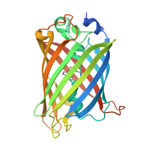
    9MUS, 9MUT, 9MUU, 9MUV

  • PubMed Abstract: 

    Genetically encoded biosensors with changes in fluorescence lifetime (as opposed to fluorescence intensity) can quantify small molecules in complex contexts, even in vivo. However, lifetime-readout sensors are poorly understood at a molecular level, complicating their development. Although there are many sensors that have fluorescence-intensity changes, there are currently only a few with fluorescence-lifetime changes. Here, we optimized two biosensors for thiol-disulfide redox (RoTq-Off and RoTq-On) with opposite changes in fluorescence lifetime in response to oxidation. Using biophysical approaches, we showed that the high-lifetime states of these sensors lock the chromophore more firmly in place than their low-lifetime states do. Two-photon fluorescence lifetime imaging of RoTq-On fused to a glutaredoxin (Grx1) enabled robust, straightforward monitoring of cytosolic glutathione redox state in acute mouse brain slices. The motional mechanism described here is probably common and may inform the design of other lifetime-readout sensors; the Grx1-RoTq-On fusion sensor will be useful for studying glutathione redox in physiology.


  • Organizational Affiliation
    • Department of Neurobiology, Harvard Medical School, Boston, MA 02115.

Macromolecule Content 

  • Total Structure Weight: 57.65 kDa 
  • Atom Count: 3,880 
  • Modeled Residue Count: 452 
  • Deposited Residue Count: 502 
  • Unique protein chains: 1

Macromolecules

Find similar proteins by:|  3D Structure
Entity ID: 1
MoleculeChains  Sequence LengthOrganismDetailsImage
Fluorescent thiol-disulfide redox biosensor
A, B
251Aequorea victoriaMutation(s): 0 
Gene Names: GFP
UniProt
Find proteins for P42212 (Aequorea victoria)
Explore P42212 
Go to UniProtKB:  P42212
Entity Groups
Sequence Clusters30% Identity50% Identity70% Identity90% Identity95% Identity100% Identity
UniProt GroupP42212
Sequence Annotations
Expand
Reference Sequence

Small Molecules

Ligands 5 Unique
IDChains Name / Formula / InChI Key2D Diagram3D Interactions
PGE

Query on PGE


Download:Ideal Coordinates CCD File
M [auth B]TRIETHYLENE GLYCOL
C6 H14 O4
ZIBGPFATKBEMQZ-UHFFFAOYSA-N
GOL

Query on GOL


Download:Ideal Coordinates CCD File
H [auth B]GLYCEROL
C3 H8 O3
PEDCQBHIVMGVHV-UHFFFAOYSA-N
EDO

Query on EDO


Download:Ideal Coordinates CCD File
C [auth A],
F [auth A]
1,2-ETHANEDIOL
C2 H6 O2
LYCAIKOWRPUZTN-UHFFFAOYSA-N
ACT

Query on ACT


Download:Ideal Coordinates CCD File
D [auth A],
E [auth A],
I [auth B],
K [auth B]
ACETATE ION
C2 H3 O2
QTBSBXVTEAMEQO-UHFFFAOYSA-M
FMT

Query on FMT


Download:Ideal Coordinates CCD File
G [auth A],
J [auth B],
L [auth B]
FORMIC ACID
C H2 O2
BDAGIHXWWSANSR-UHFFFAOYSA-N
Modified Residues  1 Unique
IDChains TypeFormula2D DiagramParent
SWG
Query on SWG
A, B
L-PEPTIDE LINKINGC16 H16 N4 O4SER, TRP, GLY

Experimental Data & Validation

Experimental Data

  • Method: X-RAY DIFFRACTION
  • Resolution: 2.20 Å
  • R-Value Free:  0.245 (Depositor), 0.247 (DCC) 
  • R-Value Work:  0.199 (Depositor), 0.201 (DCC) 
Space Group: C 2 2 21
Unit Cell:
Length ( Å )Angle ( ˚ )
a = 49.95α = 90
b = 140.84β = 90
c = 129.53γ = 90
Software Package:
Software NamePurpose
PHENIXrefinement
XDSdata reduction
XDSdata scaling

Structure Validation

View Full Validation Report


Entry History 

& Funding Information

Deposition Data


Funding OrganizationLocationGrant Number
National Institutes of Health/National Institute of General Medical Sciences (NIH/NIGMS)United StatesR01GM124038

Revision History  (Full details and data files)

  • Version 1.0: 2025-06-18
    Type: Initial release
  • Version 1.1: 2025-06-25
    Changes: Database references, Refinement description, Structure summary
  • Version 2.0: 2026-03-18
    Changes: Polymer sequence