9MT3 | pdb_00009mt3

Candida albicans Hsp90 nucleotide binding domain in complex with XL888


Experimental Data Snapshot

  • Method: X-RAY DIFFRACTION
  • Resolution: 2.02 Å
  • R-Value Free: 
    0.244 (Depositor), 0.244 (DCC) 
  • R-Value Work: 
    0.188 (Depositor), 0.188 (DCC) 
  • R-Value Observed: 
    0.193 (Depositor) 

Starting Model: experimental
View more details

wwPDB Validation 3D Report Full Report

Validation slider image for 9MT3

Ligand Structure Quality Assessment 


This is version 1.1 of the entry. See complete history

Literature

Structural Insights into Selectively Targeting Candida albicans Hsp90.

Kowalewski, M.E.Zagler, S.Redinbo, M.R.

(2025) Biochemistry 64: 2401-2411

  • DOI: https://doi.org/10.1021/acs.biochem.5c00015
  • Primary Citation Related Structures: 
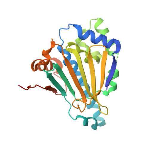
    9MSQ, 9MSU, 9MSV, 9MSX, 9MT3, 9MT9, 9MTB

  • PubMed Abstract: 

    The threat of drug-resistant pathogens continues to rise and underscores the need for new antimicrobial and antifungal strategies. Diverse chemical scaffolds have been shown with high affinity to bind the human heat-shock protein Hsp90. Orthologous proteins are present in microbial pathogens and have been shown to be particularly abundant in these organisms, suggesting they may serve as therapeutic targets. Here, we examine the potency and selectivity of human Hsp90 ligands for their capacity to bind to the nucleotide binding domain of Hsp90 from the pathogenic fungi, Candida albicans . Using a series of biochemical, structural, and fragment and in silico screening investigations, we define key chemical features that lead to effective C. albicans Hsp90 (CaHsp90) binding. We support these studies with crystal structures of five diverse human Hsp90 ligands in complex with CaHsp90, as well as the structure of this protein with a nonhydrolyzable ATP analog. We demonstrate the structural basis for the selectivity of the human Hsp90 inhibitor TAS116 for CaHsp90, features that may be exploited in the future development of improved CaHsp90 inhibitors.


  • Organizational Affiliation
    • Department of Biochemistry and Biophysics, University of North Carolina, Chapel Hill, North Carolina 27599, United States.

Macromolecule Content 

  • Total Structure Weight: 26.85 kDa 
  • Atom Count: 1,822 
  • Modeled Residue Count: 209 
  • Deposited Residue Count: 232 
  • Unique protein chains: 1

Macromolecules

Find similar proteins by:|  3D Structure
Entity ID: 1
MoleculeChains  Sequence LengthOrganismDetailsImage
Heat shock protein 90 homolog232Candida albicansMutation(s): 0 
Gene Names: HSP90CAALFM_C702030WACaJ7.0234CaO19.13868CaO19.6515
UniProt
Find proteins for P46598 (Candida albicans (strain SC5314 / ATCC MYA-2876))
Explore P46598 
Go to UniProtKB:  P46598
Entity Groups
Sequence Clusters30% Identity50% Identity70% Identity90% Identity95% Identity100% Identity
UniProt GroupP46598
Sequence Annotations
Expand
Reference Sequence

Small Molecules

Ligands 1 Unique
IDChains Name / Formula / InChI Key2D Diagram3D Interactions
99B
(Subject of Investigation/LOI)

Query on 99B


Download:Ideal Coordinates CCD File
B [auth A]5-[(2R)-butan-2-ylamino]-N-{(3-endo)-8-[5-(cyclopropylcarbonyl)pyridin-2-yl]-8-azabicyclo[3.2.1]oct-3-yl}-2-methylbenzene-1,4-dicarboxamide
C29 H37 N5 O3
LHGWWAFKVCIILM-HLRQEUIKSA-N

Experimental Data & Validation

Experimental Data

  • Method: X-RAY DIFFRACTION
  • Resolution: 2.02 Å
  • R-Value Free:  0.244 (Depositor), 0.244 (DCC) 
  • R-Value Work:  0.188 (Depositor), 0.188 (DCC) 
  • R-Value Observed: 0.193 (Depositor) 
Space Group: C 2 2 21
Unit Cell:
Length ( Å )Angle ( ˚ )
a = 81.337α = 90
b = 108.946β = 90
c = 50.735γ = 90
Software Package:
Software NamePurpose
PHENIXrefinement
PHENIXphasing
AutoProcessdata processing
Aimlessdata scaling

Structure Validation

View Full Validation Report


Ligand Structure Quality Assessment 


Entry History 

& Funding Information

Deposition Data


Funding OrganizationLocationGrant Number
National Institutes of Health/National Institute of General Medical Sciences (NIH/NIGMS)United StatesT32GM148376-01A1

Revision History  (Full details and data files)

  • Version 1.0: 2025-06-04
    Type: Initial release
  • Version 1.1: 2025-06-18
    Changes: Database references