The role of conserved elements in an active site alpha-helix of coproheme decarboxylase.
Carriuolo, A., Parrott, S., Bauer, O., Pritchett, C., Baltes, M., Phillips, R.S., Lanzilotta, W.N.(2025) J Inorg Biochem 274: 113101-113101
- PubMed: 41106177 Search on PubMed
- DOI: https://doi.org/10.1016/j.jinorgbio.2025.113101
- Primary Citation Related Structures:
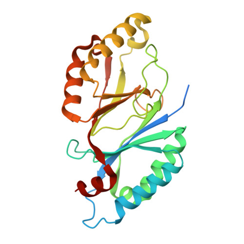
9MHQ, 9MHR - PubMed Abstract:
The final step in the coproporphyrin-dependent (CPD) branch of the heme biosynthesis pathway involves the oxidative decarboxylation of coproheme to form heme b. This reaction, catalyzed by coproheme decarboxylase (ChdC), requires two equivalents of hydrogen peroxide to complete the synthesis of one b-type heme molecule. The CPD pathway is limited to Gram-positive bacteria and some archaea, and the precise mechanism of ChdC differs between Firmicutes and Actinobacteria. These variations highlight the importance of studying ChdCs from diverse organisms. The reaction proceeds through two sequential oxidative decarboxylations via the intermediate monovinyl monopropionate deuteroheme (MMD). Previous studies suggest that MMD does not exit the active site but instead undergoes a 90-degree rotation before another equivalent of hydrogen peroxide binds and initiates the second oxidative decarboxylation. This mechanism requires a high degree of specificity to distinguish between substrate, intermediate, and final product. To further understand this selectivity, we present biochemical and structural analyses of wild-type and variant forms of ChdC from Streptomyces coelicolor (ScChdC). We hypothesize that a conserved active site element within an alpha helix contributes to porphyrin specificity/selectivity and conformation and investigate how this influences an active site loop. Our data provides new insight into the role of this loop in substrate recognition, rotation, and catalysis. The substrate selectivity model for ChdC developed in this study will inform future mechanistic investigations and provide insights into key functional interfaces, highlighting potential targets for drug development.
- Department of Biochemistry and Molecular Biology, University of Georgia, United States.
Organizational Affiliation: