9MD8 | pdb_00009md8

Hip1 complex with inhibitor #2 (Hip1-2) via Ser228


Experimental Data Snapshot

  • Method: X-RAY DIFFRACTION
  • Resolution: 2.30 Å
  • R-Value Free: 
    0.235 (Depositor), 0.236 (DCC) 
  • R-Value Work: 
    0.197 (Depositor), 0.198 (DCC) 
  • R-Value Observed: 
    0.199 (Depositor) 

Starting Model: experimental
View more details

wwPDB Validation 3D Report Full Report

Validation slider image for 9MD8

Ligand Structure Quality Assessment 


This is version 1.1 of the entry. See complete history

Literature

Discovery of highly potent alpha-keto ester-based peptidomimetic inhibitors of the Hip1 protease for the treatment of Mycobacterium tuberculosis.

Schumann, N.Shamma, F.Brooks, C.L.Johnson, S.J.Yim, M.K.Olsen, K.J.Pena, K.Karakousis, P.C.Abell, A.Goldfarb, N.E.

(2025) Eur J Med Chem Rep 15

  • DOI: https://doi.org/10.1016/j.ejmcr.2025.100311
  • Primary Citation Related Structures: 
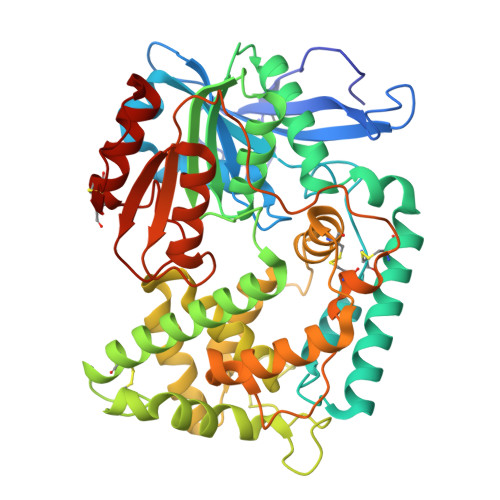
    9MD7, 9MD8

  • PubMed Abstract: 

    Mycobacterium tuberculosis (Mtb), the bacterium responsible for tuberculosis, is the leading cause of death due to a single infectious agent. Given the alarming increase in drug-resistant cases, therapeutic agents targeting novel Mtb drug targets are urgently needed. Hip1, a serine protease required for Mtb survival in macrophages and tolerance to various antibiotics, has been identified as an attractive therapeutic target. In the current study, we describe the design and synthesis of highly potent (pM range K i ) peptidomimetic α-keto ester inhibitors of Hip1. We also report the first two X-ray cocrystal structures of Hip1 bound to these compounds and describe the binding interactions in the active site of recombinant Hip1. Finally, we show that these compounds effectively reduce the intracellular bacillary burden in a macrophage model of Mtb infection.


  • Organizational Affiliation
    • Department of Chemistry, The University of Adelaide, North Terrace Campus, Adelaide, SA, 5005, Australia.

Macromolecule Content 

  • Total Structure Weight: 52.84 kDa 
  • Atom Count: 3,837 
  • Modeled Residue Count: 473 
  • Deposited Residue Count: 486 
  • Unique protein chains: 1

Macromolecules

Find similar proteins by:|  3D Structure
Entity ID: 1
MoleculeChains  Sequence LengthOrganismDetailsImage
Protease486Mycobacterium tuberculosisMutation(s): 0 
Gene Names: tapERS007688_01841
EC: 3.4 (PDB Primary Data), 3.4.14 (PDB Primary Data), 3.4.21 (UniProt)
UniProt
Find proteins for P9WHR3 (Mycobacterium tuberculosis (strain ATCC 25618 / H37Rv))
Explore P9WHR3 
Go to UniProtKB:  P9WHR3
Entity Groups
Sequence Clusters30% Identity50% Identity70% Identity90% Identity95% Identity100% Identity
UniProt GroupP9WHR3
Sequence Annotations
Expand
Reference Sequence

Small Molecules

Ligands 2 Unique
IDChains Name / Formula / InChI Key2D Diagram3D Interactions
A1BK3

Query on A1BK3


Download:Ideal Coordinates CCD File
B [auth A]N-carboxy-L-phenylalanyl-N-{(2S,3R)-3-hydroxy-4-methoxy-4-oxo-1-[(3R)-2-oxo-3,4-dihydro-2H-pyrrol-3-yl]butan-2-yl}-L-lysinamide
C25 H35 N5 O8
WFSLHCGPTMFRHH-VYJAJWGXSA-N
GOL
(Subject of Investigation/LOI)

Query on GOL


Download:Ideal Coordinates CCD File
C [auth A]GLYCEROL
C3 H8 O3
PEDCQBHIVMGVHV-UHFFFAOYSA-N

Experimental Data & Validation

Experimental Data

  • Method: X-RAY DIFFRACTION
  • Resolution: 2.30 Å
  • R-Value Free:  0.235 (Depositor), 0.236 (DCC) 
  • R-Value Work:  0.197 (Depositor), 0.198 (DCC) 
  • R-Value Observed: 0.199 (Depositor) 
Space Group: P 31 2 1
Unit Cell:
Length ( Å )Angle ( ˚ )
a = 103.725α = 90
b = 103.725β = 90
c = 127.189γ = 120
Software Package:
Software NamePurpose
PHENIXrefinement
HKL-3000data scaling

Structure Validation

View Full Validation Report


Ligand Structure Quality Assessment 


Entry History 

& Funding Information

Deposition Data


Funding OrganizationLocationGrant Number
Other privateUnited States--

Revision History  (Full details and data files)

  • Version 1.0: 2025-12-17
    Type: Initial release
  • Version 1.1: 2026-04-15
    Changes: Database references