Structural characterization of zebrafish Ngly2, an ovary-enriched acid PNGase required for egg-free glycan production.
Honda, A., Kamada, K., Seino, J., Hirayama, H., Fujihira, H., Ueki, M., Shiraki, T., Burton-Smith, R.N., Murata, K., Ishii, N., Matsuo, I., Suzuki, T.(2025) J Biological Chem 301: 110906-110906
- PubMed: 41203122 Search on PubMed
- DOI: https://doi.org/10.1016/j.jbc.2025.110906
- Primary Citation Related Structures:
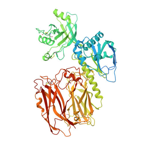
9LWG - PubMed Abstract:
Peptide:N-glycanase (PNGase) is a deglycosylating enzyme acting on asparagine(N)-linked glycans on glycoproteins. It is well established that fish possesses two PNGases with distinct properties. One is a cytosolic PNGase (NGLY1 in humans), active at neutral pH and widely conserved among eukaryotes. The other is called acid PNGase and is found in fish embryos; it is active at acidic pH and is believed to be of lysosomal origin. The gene encoding the acid PNGase has not been identified in animals, and its evolutionary distribution has remained unknown. In this study, we identified the gene encoding the acid PNGase, which we named Ngly2, in zebrafish (Danio rerio). Interestingly, zebrafish Ngly2 was found to have structural similarity with bacterial PNGase (PNGase F) and indeed appeared to share common catalytic residues, despite the fact that these two enzymes exhibit quite distinct pH profiles. The structure of zebrafish Ngly2 was determined by cryo-EM, showing that it forms homodimers and that its substrate is accommodated in the cleft between the protease-associated domain and PNGase domain, where the catalytic residues are located. Tissue distribution analysis indicated that ngly2 was almost exclusively expressed in the ovary. A zebrafish ngly2-KO line was found to be fertile, survive well, and show no overt phenotypes, although it had significantly smaller fertilized eggs. It was also revealed that ngly2 KO resulted in a substantial reduction in the level of free oligosaccharides in fertilized eggs, implying that Ngly2, not Ngly1, is responsible for the formation of most, if not all, egg-free glycans.
- Glycometabolic Biochemistry Laboratory, RIKEN Pioneering Research Institute, RIKEN, Wako, Saitama, Japan. Electronic address: akinobu.honda@riken.jp.
Organizational Affiliation: