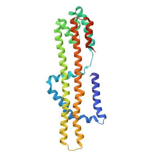
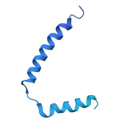
9LUB | pdb_00009lub

The chimeric flagellar motor complex between MotA1B1 from Paenibacillus sp. TCA20 and MotAB from E.coli, state 2


Experimental Data Snapshot

  • Method: ELECTRON MICROSCOPY
  • Resolution: 3.30 Å
  • Aggregation State: PARTICLE 
  • Reconstruction Method: SINGLE PARTICLE 

Starting Model: in silico
View more details

wwPDB Validation 3D Report Full Report

Validation slider image for 9LUB

This is version 1.0 of the entry. See complete history. 

Literature

Cryo-EM Structure of the Flagellar Motor Complex from Paenibacillus sp. TCA20.

Onoe, S., Nishikino, T., Kinoshita, M., Takekawa, N., Minamino, T., Imada, K., Namba, K., Kishikawa, J.I., Kato, T.

(2025) Biomolecules 15

  • DOI: https://doi.org/10.3390/biom15030435
  • Primary Citation Related Structures: 
    9LU9, 9LUB, 9LUC

  • PubMed Abstract: 

    The bacterial flagellum, a complex nanomachine composed of numerous proteins, is utilized by bacteria for swimming in various environments and plays a crucial role in their survival and infection. The flagellar motor is composed of a rotor and stator complexes, with each stator unit functioning as an ion channel that converts flow from outside of cell membrane into rotational motion. Paenibacillus sp. TCA20 was discovered in a hot spring, and a structural analysis was conducted on the stator complex using cryo-electron microscopy to elucidate its function. Two of the three structures (Classes 1 and 3) were found to have structural properties typical for other stator complexes. In contrast, in Class 2 structures, the pentamer ring of the A subunits forms a C-shape, with lauryl maltose neopentyl glycol (LMNG) bound to the periplasmic side of the interface between the A and B subunits. This interface is conserved in all stator complexes, suggesting that hydrophobic ligands and lipids can bind to this interface, a feature that could potentially be utilized in the development of novel antibiotics aimed at regulating cell motility and infection.


  • Organizational Affiliation: 
    • Institute for Protein Research, Osaka University, Suita 565-0871, Osaka, Japan.

Macromolecule Content 

  • Total Structure Weight: 213.37 kDa 
  • Atom Count: 10,113 
  • Modeled Residue Count: 1,326 
  • Deposited Residue Count: 1,958 
  • Unique protein chains: 2

Macromolecules

Find similar proteins by:|  3D Structure
Entity ID: 1
MoleculeChains  Sequence LengthOrganismDetailsImage
Flagellar motor protein MotA
A, B, C, D, E
264Paenibacillus sp. TCA20Mutation(s): 0 
Gene Names: TCA2_3717
Membrane Entity: Yes 
UniProt
Find proteins for A0AAX3N6G0 (Paenibacillus urinalis)
Explore A0AAX3N6G0 
Go to UniProtKB:  A0AAX3N6G0
Entity Groups
Sequence Clusters30% Identity50% Identity70% Identity90% Identity95% Identity100% Identity
UniProt GroupA0AAX3N6G0
Sequence Annotations
Expand
Reference Sequence
Find similar proteins by:|  3D Structure
Entity ID: 2
MoleculeChains  Sequence LengthOrganismDetailsImage
Chimeric B subunit of MotA1B1 from Paenibacillus sp. TCA20 and MotAB from E. coli,Motility protein B
F, G
319Paenibacillus sp. TCA20, Escherichia coli
This entity is chimeric
Mutation(s): 0 
Membrane Entity: Yes 
UniProt
Find proteins for P0AF06 (Escherichia coli (strain K12))
Explore P0AF06 
Go to UniProtKB:  P0AF06
Entity Groups
Sequence Clusters30% Identity50% Identity70% Identity90% Identity95% Identity100% Identity
UniProt GroupP0AF06
Sequence Annotations
Expand
Reference Sequence

Experimental Data & Validation

Experimental Data

  • Method: ELECTRON MICROSCOPY
  • Resolution: 3.30 Å
  • Aggregation State: PARTICLE 
  • Reconstruction Method: SINGLE PARTICLE 
EM Software:
TaskSoftware PackageVersion
MODEL REFINEMENTServalcat
RECONSTRUCTIONcryoSPARC3.8

Structure Validation

View Full Validation Report


Entry History 

& Funding Information

Deposition Data


Funding OrganizationLocationGrant Number
Japan Agency for Medical Research and Development (AMED)JapanJP21am0101XXX.
Japan Society for the Promotion of Science (JSPS)JapanJP23K14157

Revision History  (Full details and data files)

  • Version 1.0: 2025-04-09
    Type: Initial release