Structural basis of Ebp pilus shaft formation and anchoring in vancomycin-resistant Enterococci.
Sharma, V., Krishnan, V.(2025) FEBS J 292: 5723-5749
- PubMed: 40608565 Search on PubMed
- DOI: https://doi.org/10.1111/febs.70173
- Primary Citation Related Structures:
9LJ6, 9LKS, 9LLW, 9LR7, 9LTY, 9M00 - PubMed Abstract:
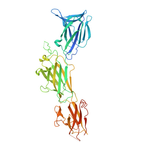
Enterococcus faecalis is an opportunistic pathogen that causes various clinically significant infections, including infective endocarditis and catheter-associated urinary tract infections. E. faecalis assembles hair-like appendages termed endocarditis and biofilm-associated pili (Ebp), crucial for adherence, colonization, biofilm formation, and virulence. The Ebp pilus comprises three pilin subunits (EbpA, EbpB, and EbpC) encoded by the ebpABC pilus operon. EbpC forms the Ebp pilus backbone decorated with EbpA and EbpB at the tip and base for adhesion and anchoring, respectively. Experimental structures are not yet available for any of the Ebp pilins. Herein, we report the crystal structures of EbpC, EbpB, and their homologs from E. faecium. The structures of EbpC and EbpB reveal four and three linearly arranged immunoglobulin-like domains, respectively. The basal pilin EbpB structure fully mimics the features of backbone pilins with a typical pilin motif and CnaB-CnaA-CnaB fold, each with an intact isopeptide bond. The rigid C-terminal fragment of the backbone pilin EbpC, containing three domains, exhibits a CnaB-CnaA-CnaB fold, each with an intradomain isopeptide bond. The flexible N-terminal domain with the CnaB fold in EbpC lacks an isopeptide bond but appears to form in a full-length structure that shows a 'beads on a string' architecture, previously unobserved for four-domain backbone pilins. Structural analysis helped us to identify residues of internal isopeptide bonds and pilin motifs harboring lysine residues responsible for intermolecular covalent links during sortase-mediated pilus assembly and propose a structural model for the Ebp pilus. This knowledge may be useful for developing structure-based approaches targeting pilins and pili in enterococcal infections.
- Laboratory of Structural Microbiology, Regional Centre for Biotechnology, NCR Biotech Science Cluster, Faridabad, India.
Organizational Affiliation: