Molecular basis of SAM-AMP synthesis and degradation in the type III-B CRISPR-Cas system.
Duan, B., Jin, X., An, X., Xiao, Y., Yang, Q., Zhao, H., Huang, Y., Wang, J., Wang, Q., Du, F., Lu, L., Sun, L., Chen, Z., Zhao, B.(2025) Nat Chem Biol
- PubMed: 41272318 Search on PubMedSearch on PubMed Central
- DOI: https://doi.org/10.1038/s41589-025-02075-z
- Primary Citation Related Structures:
9LQ5, 9LQ6, 9LQ7, 9LQ8, 9LQ9, 9LQA, 9LQB, 9LQC, 9LQD, 9LQE, 9LQF, 9LQG, 9LQH - PubMed Abstract:
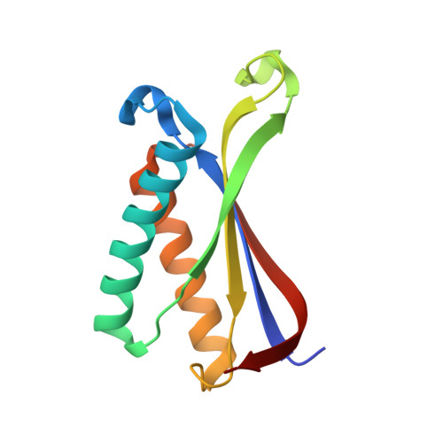
Upon sensing nonself target RNA, the CorA-associated type III-B CRISPR-Cas system catalyzes S-adenosyl methionine (SAM) and ATP to synthesize SAM-AMP, which activates the effector CorA and triggers immune responses. SAM-AMP can be degraded by NrN and SAM lyase, potentially deactivating the system. Here we find that the type III-B effector complex from Bacteroides fragilis uses a specific mechanism to recognize nonself target RNA and synthesize SAM-AMP. The 3' anti-tag of nonself target RNA induces conformational changes in the Cmr2 subunit, triggering SAM-AMP synthesis independently of the stalk loop of Cmr3 subunit. SAM-AMP binding induces NrN to transit from an open to a closed conformation, enabling hydrolysis of the 3'-5' phosphodiester bond. SAM lyase forms a triangular trimer that specifically degrades SAM-AMP into 5'-methylthioadenosine-AMP and homoserine lactone. These findings unveil unique mechanisms for SAM-AMP synthesis and degradation and provide deeper insights into the molecular basis of type III CRISPR-Cas signaling.
- Key Laboratory of Medical Molecular Virology (MOE/NHC/CAMS), Shanghai Frontiers Science Center of Pathogenic Microorganisms and Infection, Department of Medical Microbiology and Parasitology, School of Basic Medical Sciences, Shanghai Fifth People's Hospital, Shanghai Institute of Infectious Disease and Biosecurity, Institutes of Biomedical Sciences, Shanghai Medical College, Fudan University, Shanghai, China.
Organizational Affiliation: