9LJJ | pdb_00009ljj

cryo-EM structure of a nanobody bound heliorhodopsin


Experimental Data Snapshot

  • Method: ELECTRON MICROSCOPY
  • Resolution: 3.23 Å
  • Aggregation State: PARTICLE 
  • Reconstruction Method: SINGLE PARTICLE 

wwPDB Validation   3D Report Full Report


This is version 1.0 of the entry. See complete history


Literature

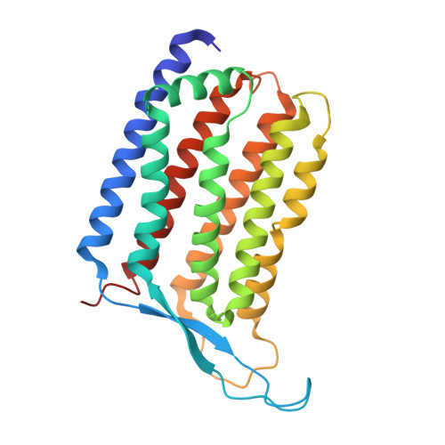
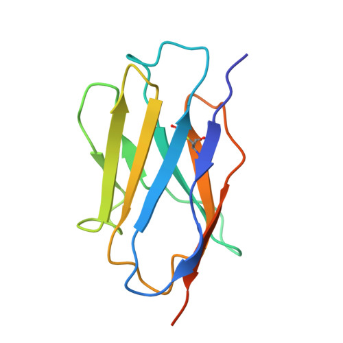
Cryo-EM structure of a nanobody-bound heliorhodopsin.

Xia, R.Sun, M.Lu, Y.Wang, N.Zhang, A.Guo, C.Xu, Z.Cai, X.He, Y.

(2025) Biochem Biophys Res Commun 750: 151398-151398

  • DOI: https://doi.org/10.1016/j.bbrc.2025.151398
  • Primary Citation of Related Structures:  
    9LJJ

  • PubMed Abstract: 

    Heliorhodopsins (HeRs) represent a distinct class of microbial rhodopsins (MRs) with an inverted membrane topology compared to other MRs. Previous structural studies have shown that HeRs lack a proton acceptor residue, and protons are never released from the protein. In this study, we present the cryo-electron microscopy (cryo-EM) structure of HeR bound to a nanobody. The structure reveals an acetate-like molecule in the Schiff base cavity (SBC) on the intracellular side of HeR under neutral condition. Structural comparisons and analyses suggest that the acetate molecule may function as a proton acceptor for the protonated retinal Schiff base (RSB) and act as a mediator for the intramolecular signaling transduction in HeR during light stimulation. These structural insights shed new light on the mechanism and function of HeR.


  • Organizational Affiliation
    • HIT Center for Life Sciences, School of Life Science and Technology, Faculty of Life Sciences and Medicine, Harbin Institute of Technology, Harbin, 150001, China.

Macromolecules
Find similar proteins by:  (by identity cutoff)  |  3D Structure
Entity ID: 1
MoleculeChains Sequence LengthOrganismDetailsImage
Heliorhodopsin
A, B
256Escherichia coliMutation(s): 0 
Membrane Entity: Yes 
UniProt
Find proteins for A0A2R4S913 (uncultured Actinomycetes bacterium)
Explore A0A2R4S913 
Go to UniProtKB:  A0A2R4S913
Entity Groups  
Sequence Clusters30% Identity50% Identity70% Identity90% Identity95% Identity100% Identity
UniProt GroupA0A2R4S913
Sequence Annotations
Expand
  • Reference Sequence
Find similar proteins by:  (by identity cutoff)  |  3D Structure
Entity ID: 2
MoleculeChains Sequence LengthOrganismDetailsImage
nanobody B4C [auth M],
D [auth N]
122Vicugna pacosMutation(s): 0 
Entity Groups  
Sequence Clusters30% Identity50% Identity70% Identity90% Identity95% Identity100% Identity
Sequence Annotations
Expand
  • Reference Sequence
Small Molecules
Ligands 4 Unique
IDChains Name / Formula / InChI Key2D Diagram3D Interactions
PX2
Query on PX2

Download Ideal Coordinates CCD File 
G [auth A]1,2-DILAUROYL-SN-GLYCERO-3-PHOSPHATE
C27 H52 O8 P
OKLASJZQBDJAPH-RUZDIDTESA-M
RET
Query on RET

Download Ideal Coordinates CCD File 
E [auth A],
J [auth B]
RETINAL
C20 H28 O
NCYCYZXNIZJOKI-OVSJKPMPSA-N
PLM
Query on PLM

Download Ideal Coordinates CCD File 
H [auth A],
I [auth A],
L [auth B]
PALMITIC ACID
C16 H32 O2
IPCSVZSSVZVIGE-UHFFFAOYSA-N
ACT
Query on ACT

Download Ideal Coordinates CCD File 
F [auth A],
K [auth B]
ACETATE ION
C2 H3 O2
QTBSBXVTEAMEQO-UHFFFAOYSA-M
Experimental Data & Validation

Experimental Data

  • Method: ELECTRON MICROSCOPY
  • Resolution: 3.23 Å
  • Aggregation State: PARTICLE 
  • Reconstruction Method: SINGLE PARTICLE 
EM Software:
TaskSoftware PackageVersion
MODEL REFINEMENTPHENIX1.18.2_3874

Structure Validation

View Full Validation Report


Entry History & Funding Information

Deposition Data

  • Released Date: 2025-02-12 
  • Deposition Author(s): He, Y., Xia, R.

Funding OrganizationLocationGrant Number
National Natural Science Foundation of China (NSFC)China32070048

Revision History  (Full details and data files)

  • Version 1.0: 2025-02-12
    Type: Initial release