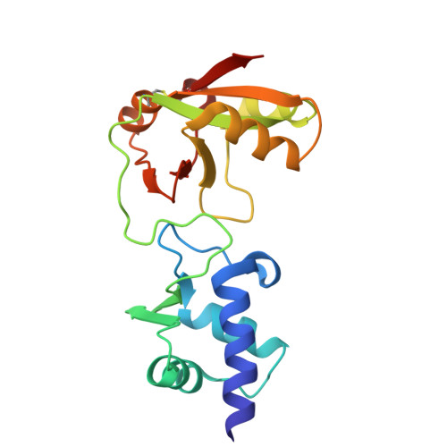
Crystal structures and snapshots along Tpt1-catalyzed phosphate transfer from nucleic acid to NAD.
Cao, C., Yang, J., Zhang, W., Chen, J., Gao, Y., Yao, Y., Zhang, Y., Li, H., Li, L., Luo, Z., Wang, C., Tang, G., Cui, R., Liu, H., Wang, Q., Huang, Z., Ma, J., Gan, J.(2025) Nat Commun 16: 10888-10888
- PubMed: 41345401 Search on PubMedSearch on PubMed Central
- DOI: https://doi.org/10.1038/s41467-025-65881-y
- Primary Citation Related Structures:
9LD3, 9LD4, 9LD6, 9LDA, 9LDC, 9LDD, 9LDE, 9LDF, 9LDG, 9LDH, 9LDI - PubMed Abstract:
Tpt1/TRPT1/KptA family proteins are evolutionarily conserved in all three domains of life. In fungi and plants, Tpt1 transfers 2'-PO 4 2- from tRNA splice junction to NAD + , which is the final step of tRNA maturation and is critical for the function of tRNA. In mammals and bacteria, Tpt1-catalyzed reaction leads to 5'-end ADP ribosylation, a reversible chemical modification of nucleic acids. Based on in vivo and in vitro biochemical studies, a two-step catalytic mechanism has been established for Tpt1-catalyzed RNA 2'-PO 4 2- transfer, including (i) the 2'-PO 4 2- attacks NAD + , releasing nicotinamide and forming a 2'-phospho-ADP-ribosylated RNA (2'-p-ADPR-RNA) intermediate; and (ii) transesterification of the ADP-ribose 2"-OH to RNA 2'-PO 4 2- , displacing the 2'-OH RNA and producing ADP-ribose-1",2"-cyclic phosphate (Appr>P). However, neither 2'-p-ADPR-RNA intermediate nor Appr>P product has been captured in any reported Tpt1 structures. Here, we report a series of crystal structures of T. kodakarensis Tpt1 (TkoTpt1), capturing the key 2'-p-ADPR-RNA intermediate. In addition, our structures also capture the 5'-p-ADPR-DNA intermediate and Appr>P product. Structural analysis and in vitro catalytic assays revealed that TkoTpt1 utilizes similar mechanism in 2'-PO 4 2- and 5'-PO 4 2- transfer. In conclusion, our structures reaffirm the catalytic mechanism of Tpt1-catalyzed phosphate transfer.
- State Key Laboratory of Genetics and Development of Complex Phenotypes, Department of Biochemistry and Biophysics, School of Life Sciences, Fudan University, Shanghai, China.
Organizational Affiliation: