Calcium dissociation with carbonate ions from Pf-SCP, sarcoplasmic calcium-binding protein in Pinctada fucata, contributes to calcium mineralization for shell formation.
Namikawa, Y., Zhu, L., Lu, P., Nagata, K., Suzuki, M.(2025) Protein Sci 34: e70336-e70336
- PubMed: 41123418 Search on PubMedSearch on PubMed Central
- DOI: https://doi.org/10.1002/pro.70336
- Primary Citation Related Structures:
9L5M - PubMed Abstract:
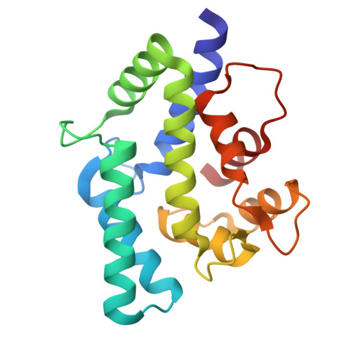
Pf-SCP is an EF-hand protein identified in Pinctada fucata that is responsible for calcium transport and concentration in the mantle for shell formation. Previous studies have reported the calcium-binding properties of the EF-hand domains and the localization of Pf-SCP. To understand the calcification from Pf-SCP as a source of calcium, the dissociation of calcium from Pf-SCP must be investigated. However, calcium dissociation from EF-hand proteins, particularly in the presence of carbonate ions, remains poorly understood. In this study, we demonstrated that calcium dissociation from Pf-SCP was induced by carbonate ions using the fluorescence spectra of Pf-SCP, and this was followed by the synthesis of calcium carbonate that was characterized using scanning electron microscope-energy dispersive X-ray spectrometry (SEM-EDS). To gain insight into the calcium dissociation of Pf-SCP at the atomic level, we conducted molecular dynamics simulations using a multi-state ion model for calcium ions. The proposed mechanism of calcium dissociation in Pf-SCP is as follows: Water molecules first replace the amino acids in the EF-hand domain to coordinate calcium ions. Next, the carbonate ions bind to the calcium ions, decreasing the binding affinity of the EF-hand domains for the calcium ions. Finally, the calcium ions detach from the EF-hand, forming a complex with water molecules and carbonate ions. These findings provide a detailed understanding of the structural dynamics of calcium dissociation and the biomineralization mechanism in P. fucata, particularly in relation to the mantle calcification process.
- Department of Applied Biological Chemistry, Graduate School of Agricultural and Life Sciences, The University of Tokyo, Tokyo, Japan.
Organizational Affiliation: