9L3V | pdb_00009l3v

structure of WEEV strain 71V1658 virus-like particle(3-fold region)


Experimental Data Snapshot

  • Method: ELECTRON MICROSCOPY
  • Resolution: 2.53 Å
  • Aggregation State: PARTICLE 
  • Reconstruction Method: SINGLE PARTICLE 

wwPDB Validation   3D Report Full Report


This is version 1.0 of the entry. See complete history


Literature

Structural basis for engagement of Western Equine Encephalitis Virus with the PCDH10 receptor.

Liang, S.Yang, Y.Liu, Y.Xu, Z.Hou, J.Li, D.Zhao, L.Hu, C.Liu, X.Rao, Z.Wang, Y.Lou, Z.

(2025) Nat Commun 16: 6290-6290

  • DOI: https://doi.org/10.1038/s41467-025-61659-4
  • Primary Citation of Related Structures:  
    9L3V, 9L41

  • PubMed Abstract: 

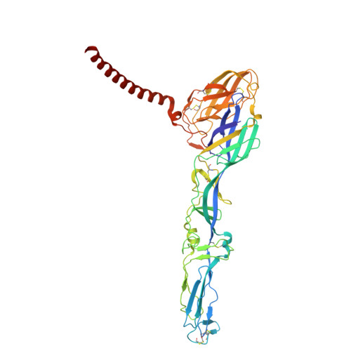
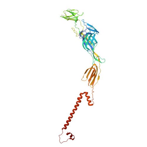
    PCDH10 is a newly identified general receptor for Western equine encephalitis virus (WEEV) members, a group of encephalitic alphaviruses that cause severe diseases in humans and equids. While WEEV typically binds PCDH10 as a receptor, nonpathogenic strains have evolved to lose mammalian PCDH10 binding, retaining only avian PCDH10 affinity. Virulent strains also engage VLDLR and ApoER2 as alternative receptors. Here, we determine the structure of WEEV strain 71V1658 virus-like particles (VLPs) in complex with human PCDH10 extracellular cadherin repeats 1-2 (EC1-EC2) by cryo-electron microscopy at 2.99 Å resolution. EC1 inserts into a cleft clamped by two adjacent E2-E1 heterodimers within a single trimeric spike, whereas EC2 maintains no contact with the WEEV VLP. Mutagenesis studies elucidate the impacts of the interacting residues on PCDH10. And residue 153 of E2 is crucial for PCDH10 binding, and the E2 Q153L mutation observes in the nonpathogenic strain Imperial-181 restores its ability to bind to PCDH10. Moreover, the arginine residue at position 89 on avian PCDH10 is essential for its interaction with strain Imperial-181. These results advance our understanding of receptor recognition by alphaviruses and the shift in receptor usage, providing insights for the development of antiviral therapies.


  • Organizational Affiliation
    • School of Basic Medical Sciences, Tsinghua University, Beijing, China.

Macromolecules
Find similar proteins by:  (by identity cutoff)  |  3D Structure
Entity ID: 1
MoleculeChains Sequence LengthOrganismDetailsImage
Structural polyprotein
A, C, E
439Western equine encephalitis virusMutation(s): 0 
Membrane Entity: Yes 
UniProt
Find proteins for P13897 (Western equine encephalitis virus)
Explore P13897 
Go to UniProtKB:  P13897
Entity Groups  
Sequence Clusters30% Identity50% Identity70% Identity90% Identity95% Identity100% Identity
UniProt GroupP13897
Sequence Annotations
Expand
  • Reference Sequence
Find similar proteins by:  (by identity cutoff)  |  3D Structure
Entity ID: 2
MoleculeChains Sequence LengthOrganismDetailsImage
Structural polyprotein
B, D, F
418Western equine encephalitis virusMutation(s): 0 
Membrane Entity: Yes 
UniProt
Find proteins for P13897 (Western equine encephalitis virus)
Explore P13897 
Go to UniProtKB:  P13897
Entity Groups  
Sequence Clusters30% Identity50% Identity70% Identity90% Identity95% Identity100% Identity
UniProt GroupP13897
Sequence Annotations
Expand
  • Reference Sequence
Experimental Data & Validation

Experimental Data

  • Method: ELECTRON MICROSCOPY
  • Resolution: 2.53 Å
  • Aggregation State: PARTICLE 
  • Reconstruction Method: SINGLE PARTICLE 
EM Software:
TaskSoftware PackageVersion
MODEL REFINEMENTPHENIX1.20.1_4487
RECONSTRUCTIONcryoSPARC

Structure Validation

View Full Validation Report


Entry History & Funding Information

Deposition Data


Funding OrganizationLocationGrant Number
National Natural Science Foundation of China (NSFC)China--

Revision History  (Full details and data files)

  • Version 1.0: 2025-08-27
    Type: Initial release