9L14 | pdb_00009l14

Crystal structure of the monobody CL-1 in complex with the Escherichia coli adenylate kinase


Experimental Data Snapshot

  • Method: X-RAY DIFFRACTION
  • Resolution: 1.86 Å
  • R-Value Free: 
    0.235 (Depositor), 0.235 (DCC) 
  • R-Value Work: 
    0.192 (Depositor), 0.191 (DCC) 
  • R-Value Observed: 
    0.194 (Depositor) 

Starting Model: experimental
View more details

wwPDB Validation   3D Report Full Report


Ligand Structure Quality Assessment 


This is version 1.0 of the entry. See complete history


Literature

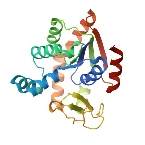
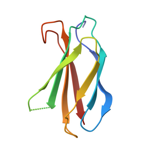
Binding mechanism of adenylate kinase-specific monobodies.

Nakamura, I.Amesaka, H.Nagao, S.Orito, N.Negi, S.Tanaka, S.I.Matsuo, T.

(2025) FEBS Lett 

  • DOI: https://doi.org/10.1002/1873-3468.70076
  • Primary Citation of Related Structures:  
    9L14

  • PubMed Abstract: 

    Monobodies are synthetic antibody-mimetic proteins that regulate enzyme functions through protein-protein interactions. In this study, we investigated the binding mechanisms of monobodies to adenylate kinase (Adk). Calorimetric and X-ray crystallographic analyses revealed that CL-1, a monobody specific for the CLOSED form of Adk, binds to the CORE domain of Adk in an enthalpy-driven manner, forming several hydrogen bonds and a cation-π interaction at the protein interface, without perturbing the Adk backbone. In contrast, OP-4, an OPEN-form-specific monobody, exhibited entropy-driven binding. 1 H- 15 N 2D nuclear magnetic resonance (NMR), 31 P-NMR, and calorimetric studies revealed conformational perturbations to Adk by OP-4, while substrate access remained intact. The different thermodynamic and structural effects between the monobodies highlight the diverse binding mechanisms among monobodies.


  • Organizational Affiliation

    Division of Materials Science, Nara Institute of Science and Technology (NAIST), Ikoma, Nara, Japan.


Macromolecules
Find similar proteins by:  (by identity cutoff)  |  3D Structure
Entity ID: 1
MoleculeChains Sequence LengthOrganismDetailsImage
Adenylate kinase214Escherichia coli K-12Mutation(s): 0 
Gene Names: adkdnaWplsAb0474JW0463
EC: 2.7.4.3
UniProt
Find proteins for P69441 (Escherichia coli (strain K12))
Explore P69441 
Go to UniProtKB:  P69441
Entity Groups  
Sequence Clusters30% Identity50% Identity70% Identity90% Identity95% Identity100% Identity
UniProt GroupP69441
Sequence Annotations
Expand
  • Reference Sequence
Find similar proteins by:  (by identity cutoff)  |  3D Structure
Entity ID: 2
MoleculeChains Sequence LengthOrganismDetailsImage
Monobody CL-191synthetic constructMutation(s): 0 
Entity Groups  
Sequence Clusters30% Identity50% Identity70% Identity90% Identity95% Identity100% Identity
Sequence Annotations
Expand
  • Reference Sequence
Experimental Data & Validation

Experimental Data

  • Method: X-RAY DIFFRACTION
  • Resolution: 1.86 Å
  • R-Value Free:  0.235 (Depositor), 0.235 (DCC) 
  • R-Value Work:  0.192 (Depositor), 0.191 (DCC) 
  • R-Value Observed: 0.194 (Depositor) 
Space Group: P 61
Unit Cell:
Length ( Å )Angle ( ˚ )
a = 63.53α = 90
b = 63.53β = 90
c = 128.91γ = 120
Software Package:
Software NamePurpose
PHENIXrefinement
XDSdata reduction
XDSdata scaling
PHENIXphasing

Structure Validation

View Full Validation Report


Ligand Structure Quality Assessment 


Entry History & Funding Information

Deposition Data


Funding OrganizationLocationGrant Number
Japan Society for the Promotion of Science (JSPS)JapanJP22K05316

Revision History  (Full details and data files)

  • Version 1.0: 2025-06-04
    Type: Initial release