Structural insights into heterohexameric assembly of epilepsy-related ligand-receptor complex LGI1-ADAM22.
Yamaguchi, T., Okatsu, K., Kubota, M., Mitsumori, A., Yamagata, A., Fukata, Y., Fukata, M., Shibata, M., Fukai, S.(2025) Elife 14
- PubMed: 40601686 Search on PubMedSearch on PubMed Central
- DOI: https://doi.org/10.7554/eLife.105918
- Primary Citation Related Structures:
9KZC, 9KZT - PubMed Abstract:
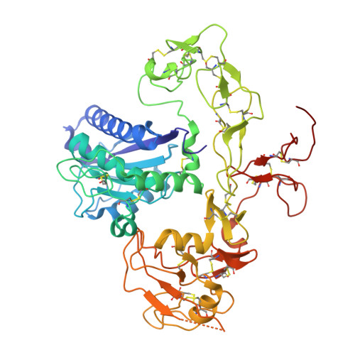
Leucine-rich glioma-inactivated 1 protein (LGI1) is a secreted neuronal protein consisting of the N-terminal leucine-rich repeat (LRR) and C-terminal epitempin-repeat (EPTP) domains. LGI1 is linked to epilepsy, a neurological disorder that can be caused by genetic mutations of genes regulating neuronal excitability (e.g. voltage- or ligand-gated ion channels). ADAM22 is a membrane receptor that binds to LGI1 extracellularly and interacts with AMPA-type glutamate receptors via PSD-95 intracellularly to maintain normal synaptic signal transmission. Structural analysis of the LGI1-ADAM22 complex is important for understanding the molecular mechanism of epileptogenesis and developing new therapies against epilepsy. We previously reported the crystal structure of a 2:2 complex consisting of two molecules of LGI1 and two molecules of the ADAM22 ectodomain (ECD), which is suggested to bridge neurons across the synaptic cleft. On the other hand, multiangle light scattering, small-angle X-ray scattering, and cryo-electron microscopy (cryo-EM) analyses have suggested the existence of a 3:3 complex consisting of three molecules of LGI1 and three molecules of ADAM22. In the previous cryo-EM analysis, many observed particles were in a dissociated state, making it difficult to determine the three-dimensional (3D) structure of the 3:3 complex. In this study, we stabilized the 3:3 LGI1-ADAM22 ECD complex using chemical cross-linking and determined the cryo-EM structures of the LGI1 LRR -LGI1 EPTP -ADAM22 ECD and 3:3 LGI1-ADAM22 ECD complexes at 2.78 Å and 3.79 Å resolutions, respectively. Furthermore, high-speed atomic force microscopy (HS-AFM) visualized the structural features and flexibility of the 3:3 LGI1-ADAM22 ECD complex in solution. We discuss new insights into the interaction modes of the LGI1-ADAM22 higher-order complex and the structural properties of the 3:3 LGI1-ADAM22 complex.
- Department of Chemistry, Graduate School of Science, Kyoto University, Kyoto, Japan.
Organizational Affiliation: