


Dihydrofolate reductase binding to NADPH and trimethoprim-tetramethylrhodamine
Zhang, K.C., Chen, Z.X.To be published.
Experimental Data Snapshot
Starting Model: experimental
View more details
Entity ID: 1 | |||||
|---|---|---|---|---|---|
| Molecule | Chains | Sequence Length | Organism | Details | Image |
| Dihydrofolate reductase | A [auth B], B [auth A] | 159 | Escherichia coli K-12 | Mutation(s): 0 Gene Names: folA, tmrA, b0048, JW0047 EC: 1.5.1.3 | 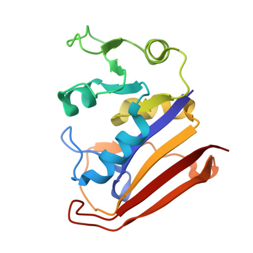 |
UniProt | |||||
Entity Groups | |||||
| Sequence Clusters | 30% Identity50% Identity70% Identity90% Identity95% Identity100% Identity | ||||
| UniProt Group | P0ABQ4 | ||||
Sequence AnnotationsExpand | |||||
Reference Sequence | |||||
| Ligands 3 Unique | |||||
|---|---|---|---|---|---|
| ID | Chains | Name / Formula / InChI Key | 2D Diagram | 3D Interactions | |
| NDP Download:Ideal Coordinates CCD File | C [auth B], H [auth A] | NADPH DIHYDRO-NICOTINAMIDE-ADENINE-DINUCLEOTIDE PHOSPHATE C21 H30 N7 O17 P3 ACFIXJIJDZMPPO-NNYOXOHSSA-N |  | ||
| A1EHX Download:Ideal Coordinates CCD File | D [auth B], I [auth A] | [9-[5-[2-[4-[[2,4-bis(azanyl)pyrimidin-5-yl]methyl]-2,6-dimethoxy-phenoxy]ethylcarbamoyl]-2-carboxy-phenyl]-6-(dimethylamino)xanthen-3-ylidene]-dimethyl-azanium C40 H42 N7 O7 JYRINKWAGFHPTL-UHFFFAOYSA-O |  | ||
| MG Download:Ideal Coordinates CCD File | E [auth B] F [auth B] G [auth B] J [auth A] K [auth A] | MAGNESIUM ION Mg JLVVSXFLKOJNIY-UHFFFAOYSA-N |  | ||
| Length ( Å ) | Angle ( ˚ ) |
|---|---|
| a = 79.995 | α = 90 |
| b = 79.995 | β = 90 |
| c = 106.073 | γ = 120 |
| Software Name | Purpose |
|---|---|
| PHENIX | refinement |
| HKL-2000 | data reduction |
| HKL-2000 | data scaling |
| PHENIX | phasing |
| Funding Organization | Location | Grant Number |
|---|---|---|
| Chinese Scholarship Council | China | 31971375 |


