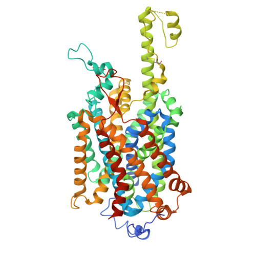
9KXT | pdb_00009kxt

Structure of human B0AT1-ACE2 complex with compound1


Experimental Data Snapshot

  • Method: ELECTRON MICROSCOPY
  • Resolution: 2.70 Å
  • Aggregation State: PARTICLE 
  • Reconstruction Method: SINGLE PARTICLE 

wwPDB Validation 3D Report Full Report

Validation slider image for 9KXT

This is version 1.1 of the entry. See complete history

Literature

Structure-guided development of a potent human B 0 AT1 inhibitor effective in a mouse model of phenylketonuria.

Imazu, T.Akashi, T.Hiraizumi, M.Inui, Y.Sasaki, W.Takahashi, T.Todoroki, H.Kumanomidou, T.Yamada, K.Fujikawa, N.Hisano, H.Asada, H.Kusakizako, T.Nishizawa, T.Iwata, S.Nureki, O.Miyaguchi, I.

(2026) Commun Biol 

  • DOI: https://doi.org/10.1038/s42003-026-10535-y
  • Primary Citation Related Structures: 
    9KXT, 9KXU, 9KXV, 9KXW, 9KXX, 9KXY, 9KXZ, 9KY0, 9KY1, 9LSZ

  • PubMed Abstract: 

    B 0 AT1 (SLC6A19) is a neutral amino acid transporter mediating intestinal absorption and renal reuptake of amino acids, including phenylalanine (Phe). Inhibiting B 0 AT1 enhances Phe excretion, offering a therapeutic strategy for phenylketonuria (PKU). Using cryo-EM, we determined human B 0 AT1 structures in outward- and inward-open states, revealing an allosteric pocket ~17 Å from the substrate site that is present in the outward-open conformation and has been previously reported. Structure-guided inhibitor design targeting this pocket produced a potent B 0 AT1 inhibitor that locks the transporter in an outward-occluded state and blocks transport. The higher-resolution structures reveal detailed interactions at the binding site, including water-mediated coordination and conformational changes around Leu52. This inhibitor exhibited submicromolar IC 50 against both human and mouse B 0 AT1, and oral administration in PKU model mice increased urinary Phe and reduced plasma Phe levels. These findings provide structural insight into allosteric inhibition of B 0 AT1 and establish a framework for the rational optimization of inhibitors targeting conformationally dynamic allosteric sites in SLC6-family transporters.


  • Organizational Affiliation
    • Neuro Science Unit Sohyaku Innovative Research Division, Mitsubishi Tanabe Pharma Co., Ltd., Yokohama, Japan. 0389158@mt-pharma.co.jp.

Macromolecule Content 

  • Total Structure Weight: 166.91 kDa 
  • Atom Count: 5,019 
  • Modeled Residue Count: 625 
  • Deposited Residue Count: 1,460 
  • Unique protein chains: 2

Macromolecules

Find similar proteins by:|  3D Structure
Entity ID: 1
MoleculeChains  Sequence LengthOrganismDetailsImage
Sodium-dependent neutral amino acid transporter B(0)AT1654Homo sapiensMutation(s): 0 
Gene Names: SLC6A19B0AT1
UniProt & NIH Common Fund Data Resources
Find proteins for Q695T7 (Homo sapiens)
Explore Q695T7 
Go to UniProtKB:  Q695T7
PHAROS:  Q695T7
GTEx:  ENSG00000174358 
Entity Groups
Sequence Clusters30% Identity50% Identity70% Identity90% Identity95% Identity100% Identity
UniProt GroupQ695T7
Glycosylation
Glycosylation Sites: 2Go to GlyGen: Q695T7-1
Sequence Annotations
Expand
Reference Sequence
Find similar proteins by:|  3D Structure
Entity ID: 2
MoleculeChains  Sequence LengthOrganismDetailsImage
Angiotensin-converting enzyme 2806Homo sapiensMutation(s): 0 
Gene Names: ACE2UNQ868/PRO1885
EC: 3.4.17.23 (PDB Primary Data), 3.4.17 (PDB Primary Data)
UniProt & NIH Common Fund Data Resources
Find proteins for Q9BYF1 (Homo sapiens)
Explore Q9BYF1 
Go to UniProtKB:  Q9BYF1
PHAROS:  Q9BYF1
GTEx:  ENSG00000130234 
Entity Groups
Sequence Clusters30% Identity50% Identity70% Identity90% Identity95% Identity100% Identity
UniProt GroupQ9BYF1
Sequence Annotations
Expand
Reference Sequence

Oligosaccharides

Help  
Entity ID: 3
MoleculeChains Length2D Diagram GlycosylationD Interactions
2-acetamido-2-deoxy-beta-D-glucopyranose-(1-4)-2-acetamido-2-deoxy-beta-D-glucopyranose
C
2N-Glycosylation
Glycosylation Resources
GlyTouCan: G42666HT
GlyCosmos: G42666HT
GlyGen: G42666HT

Small Molecules

Ligands 2 Unique
IDChains Name / Formula / InChI Key2D Diagram3D Interactions
A1L6U
(Subject of Investigation/LOI)

Query on A1L6U


Download:Ideal Coordinates CCD File
D [auth A]3-(3-bromophenyl)-~{N}-ethyl-propanamide
C11 H14 Br N O
UGAKXJNKHSOXDT-UHFFFAOYSA-N
NAG

Query on NAG


Download:Ideal Coordinates CCD File
E [auth A]2-acetamido-2-deoxy-beta-D-glucopyranose
C8 H15 N O6
OVRNDRQMDRJTHS-FMDGEEDCSA-N

Experimental Data & Validation

Experimental Data

  • Method: ELECTRON MICROSCOPY
  • Resolution: 2.70 Å
  • Aggregation State: PARTICLE 
  • Reconstruction Method: SINGLE PARTICLE 
EM Software:
TaskSoftware PackageVersion
MODEL REFINEMENTREFMAC5.8.0267

Structure Validation

View Full Validation Report


Entry History 

& Funding Information

Deposition Data


Funding OrganizationLocationGrant Number
Not funded--

Revision History  (Full details and data files)

  • Version 1.0: 2025-12-10
    Type: Initial release
  • Version 1.1: 2026-09-02
    Changes: Data collection, Database references