9KUV | pdb_00009kuv

Mechanistic insights into the versatile stoichiometry and biased signaling of the apelin receptor-arrestin complex


Experimental Data Snapshot

  • Method: ELECTRON MICROSCOPY
  • Resolution: 3.21 Å
  • Aggregation State: PARTICLE 
  • Reconstruction Method: SINGLE PARTICLE 

wwPDB Validation   3D Report Full Report


This is version 1.0 of the entry. See complete history


Literature

Mechanistic insights into the versatile stoichiometry and biased signaling of the apelin receptor-arrestin complex.

Yue, Y.Xu, C.Wu, L.Na, M.Xu, K.Chen, X.Song, Y.Weng, S.Xu, L.Li, F.Lin, X.Wang, A.Liu, J.Xu, F.

(2025) Nat Commun 16: 7403-7403

  • DOI: https://doi.org/10.1038/s41467-025-62870-z
  • Primary Citation of Related Structures:  
    9KUV, 9KUW, 9KUX

  • PubMed Abstract: 

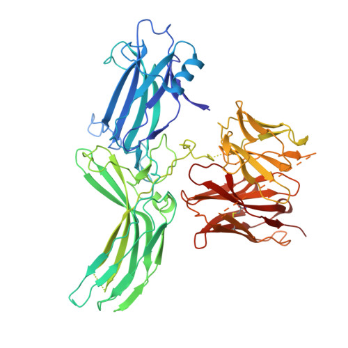
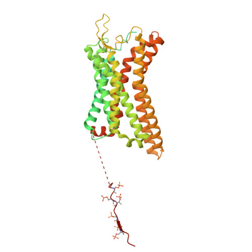
    The apelin receptor (APJR) plays a pivotal role in regulating cardiovascular and metabolic health 1,2 . Understanding the mechanisms of biased agonism at APJR is crucial for drug discovery, as stimulation of the β-arrestin pathway may lead to some adverse effects 3 . Structural analyses of APJR-Gi complexes have clarified the structural basis of receptor dimerization and activation 4,5 , yet the absence of structural data on APJR-arrestin complexes has impeded a comprehensive understanding of APJR stoichiometry in the dual signaling pathways and biased agonism. Here, we present APJR-β-arrestin1 structures bound to a clinical drug analog, revealing 2:2 and 2:1 stoichiometries associated with differential β-arrestin recruitment. Through comparison of the two transducer-coupled APJR structures bound to the same ligand, we identify key residues and motifs crucial for directing biased signaling. These findings highlight APJR's versatile stoichiometry in coupling with β-arrestin and Gi proteins, establishing a framework for understanding biased agonism and guiding the development of therapeutics.


  • Organizational Affiliation
    • iHuman Institute, ShanghaiTech University, Shanghai, China.

Macromolecules
Find similar proteins by:  (by identity cutoff)  |  3D Structure
Entity ID: 1
MoleculeChains Sequence LengthOrganismDetailsImage
Beta-arrestin-scFv30621Homo sapiensMutation(s): 0 
Entity Groups  
Sequence Clusters30% Identity50% Identity70% Identity90% Identity95% Identity100% Identity
Sequence Annotations
Expand
  • Reference Sequence
Find similar proteins by:  (by identity cutoff)  |  3D Structure
Entity ID: 2
MoleculeChains Sequence LengthOrganismDetailsImage
Apelin ReceptorB [auth R]506Homo sapiensMutation(s): 0 
UniProt & NIH Common Fund Data Resources
Find proteins for P35414 (Homo sapiens)
Explore P35414 
Go to UniProtKB:  P35414
PHAROS:  P35414
GTEx:  ENSG00000134817 
Entity Groups  
Sequence Clusters30% Identity50% Identity70% Identity90% Identity95% Identity100% Identity
UniProt GroupP35414
Sequence Annotations
Expand
  • Reference Sequence
Small Molecules
Ligands 2 Unique
IDChains Name / Formula / InChI Key2D Diagram3D Interactions
A1L6R
Query on A1L6R

Download Ideal Coordinates CCD File 
C [auth R](1~{S},2~{S})-~{N}-[4-(2,6-dimethoxyphenyl)-5-(6-methylpyridin-2-yl)-1,2,4-triazol-3-yl]-1-(5-methylpyrimidin-2-yl)-1-oxidanyl-propane-2-sulfonamide
C24 H27 N7 O5 S
HNGYVRSMVVCWGF-HRAATJIYSA-N
CLR
Query on CLR

Download Ideal Coordinates CCD File 
D [auth R]CHOLESTEROL
C27 H46 O
HVYWMOMLDIMFJA-DPAQBDIFSA-N
Modified Residues  2 Unique
IDChains TypeFormula2D DiagramParent
SEP
Query on SEP
B [auth R]L-PEPTIDE LINKINGC3 H8 N O6 PSER
TPO
Query on TPO
B [auth R]L-PEPTIDE LINKINGC4 H10 N O6 PTHR
Experimental Data & Validation

Experimental Data

  • Method: ELECTRON MICROSCOPY
  • Resolution: 3.21 Å
  • Aggregation State: PARTICLE 
  • Reconstruction Method: SINGLE PARTICLE 
EM Software:
TaskSoftware PackageVersion
MODEL REFINEMENTPHENIX1.20.1_4487
RECONSTRUCTIONcryoSPARC

Structure Validation

View Full Validation Report


Entry History & Funding Information

Deposition Data


Funding OrganizationLocationGrant Number
Not funded--

Revision History  (Full details and data files)

  • Version 1.0: 2025-09-17
    Type: Initial release