Download Mendeley
Structural and functional investigation of DinG containing a 3'-5' exonuclease domain.
Gao, T., Hao, W., Gao, J., Sun, Y., Sun, Y., Yang, J., Cheng, K.(2025) mBio 16: e0088425-e0088425
- PubMed: 40586552 Search on PubMedSearch on PubMed Central
- DOI: https://doi.org/10.1128/mbio.00884-25
- Primary Citation Related Structures:
8ZEF, 9II8, 9KQH - PubMed Abstract:
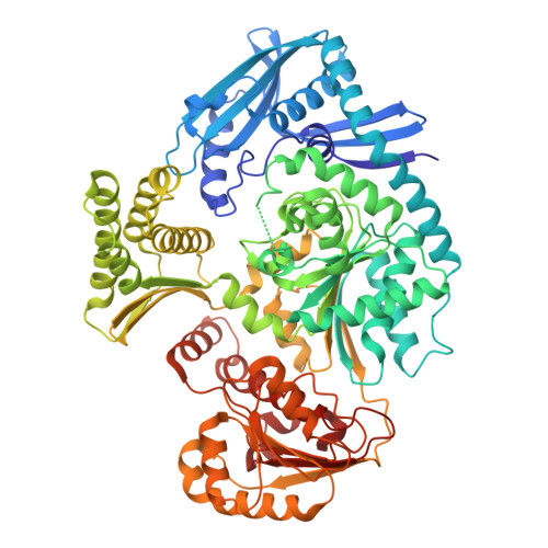
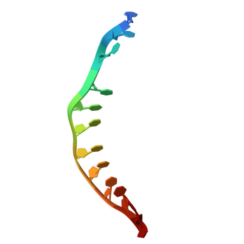
Damage-inducible gene G (DinG), a bacterial homolog of SF2 helicase, has been extensively studied in Escherichia coli . However, the structural and functional characteristics of DinG homologs fused with an N-terminal 3'-5' exonuclease domain, such as Staphylococcus aureus DinG (SaDinG), remain unexplored. In this study, we demonstrate that SaDinG possesses 3'-5' exonuclease activity and exhibits 5'-3' helicase activity on diverse DNA substrates, including splayed duplexes, 5'-overhangs, double flaps, bubbles, and gapped duplexes, resolving prior ambiguities about its biochemical functions. Intriguingly, both enzymatic activities were inhibited by elevated ATP concentrations, suggesting a potential ATP-dependent regulatory mechanism in vivo . We determined the crystal structures of SaDinG bound to ssDNA at ~3.2 Å resolution and identified key residues essential for its helicase and exonuclease activities through mutational analysis. Phenotypic studies revealed that a SaDinG deletion mutant exhibited heightened sensitivity to DNA crosslinking agents (mitomycin C and formaldehyde) but retained wild-type susceptibility to other DNA-damaging compounds. Complementation with either nuclease-dead or helicase-dead variants failed to restore crosslink resistance, indicating that both activities are indispensable for DNA crosslink repair. These results support a model in which SaDinG functions as a coordinated nuclease-helicase machine specifically adapted for DNA crosslink repair, with its dual enzymatic activities being tightly regulated by physiological ATP concentrations. DNA helicases and exonucleases play essential roles in genome maintenance; however, little is known about bacterial helicase-exonuclease fusion proteins. This study examines DNA helicases and exonucleases that play essential roles in genome maintenance; however, little is known about bacterial helicase-exonuclease fusion proteins. This study provides the first structural and functional characterization of Staphylococcus aureus DinG (SaDinG), a unique enzyme that combines 5'-3' helicase and 3'-5' exonuclease activities. Our findings resolve previous uncertainties about SaDinG's function and reveal an ATP-dependent regulatory mechanism that modulates its activity. Additionally, we demonstrate that SaDinG is critical for bacterial resistance to DNA crosslinking agents. These insights not only expand our understanding of bacterial DNA repair but also suggest potential avenues for targeting DinG-like enzymes in antimicrobial strategies. Given the growing concerns over antibiotic resistance, understanding how bacteria maintain genome integrity under stress conditions is crucial. This work lays the foundation for further exploration of bacterial helicase-exonuclease systems and their role in genome stability and adaptive survival.
- Zhejiang Key Laboratory of Medical Epigenetics, Department of Immunology and Pathogen Biology, School of Basic Medical Sciences, Affiliated Hospital of Hangzhou Normal University, Hangzhou Normal University, Hangzhou, Zhejiang, China.
Organizational Affiliation: