9KK2 | pdb_00009kk2

Cryo-EM structure of the Retron-Eco7 complex (state 5)


Experimental Data Snapshot

  • Method: ELECTRON MICROSCOPY
  • Resolution: 2.58 Å
  • Aggregation State: PARTICLE 
  • Reconstruction Method: SINGLE PARTICLE 

wwPDB Validation   3D Report Full Report


This is version 1.1 of the entry. See complete history


Literature

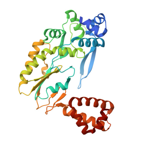
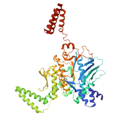
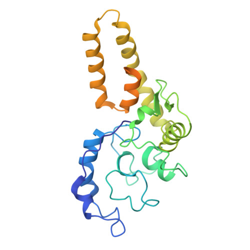
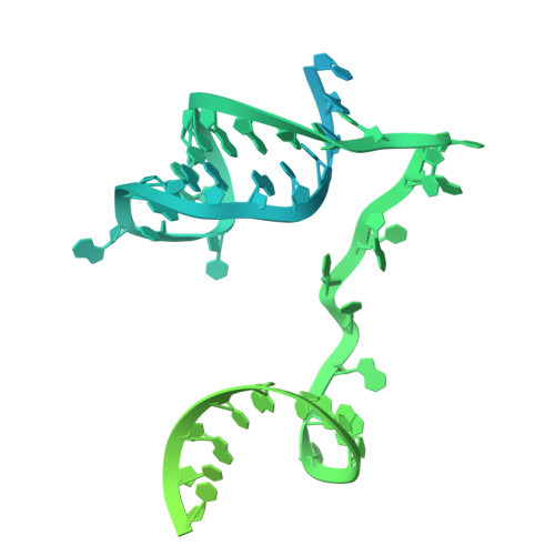
Structural mechanism of the Retron-Eco7 anti-phage defense system.

Ishikawa, J.Yoneyama, K.Azam, A.H.Nagao, A.Mitsuda, Y.Nakazaki, R.Chihara, K.Hiraizumi, M.Yamashita, K.Suzuki, T.Kiga, K.Nishimasu, H.

(2025) Nat Commun 16: 10821-10821

  • DOI: https://doi.org/10.1038/s41467-025-66589-9
  • Primary Citation of Related Structures:  
    9KJX, 9KJY, 9KJZ, 9KK1, 9KK2

  • PubMed Abstract: 

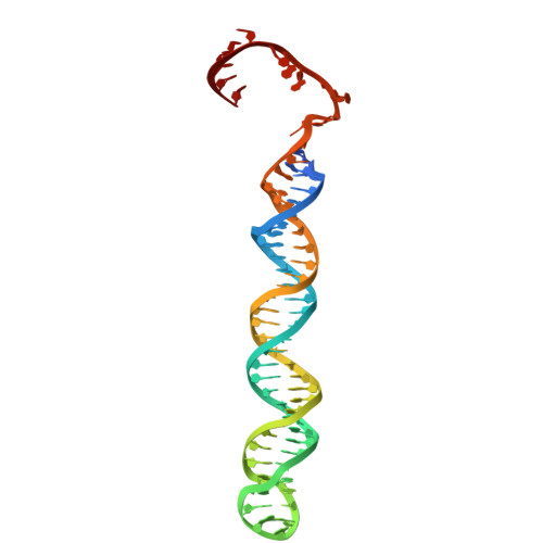
    Retrons are prokaryotic genetic elements involved in anti-phage defense and consist of a non-coding RNA, a reverse transcriptase (RT), and various effector proteins. Retron-Eco7 (previously known as Retron-Ec78) from Escherichia coli encodes two effector proteins (the PtuA ATPase and the PtuB nuclease) and degrades the host tRNA Tyr upon phage infection, thereby protecting host cells against invading phages. However, its defense mechanism remains elusive. Here, we report the cryo-electron microscopy (cryo-EM) structures of the Retron-Eco7 complex, comprising the RT, multicopy single-stranded DNA (msDNA), PtuA, and PtuB. The Retron-Eco7 structures reveal that the RT-msDNA complex associates with two PtuA-PtuB complexes, potentially inhibiting their nuclease activity and suppressing bacterial growth arrest prior to phage infection. Furthermore, the phage-encoded D15 nuclease acts as a trigger for the Retron-Eco7 system and cleaves the msDNA bound to the complex, facilitating the dissociation of PtuA-PtuB from RT-msDNA. Our data indicate that msDNA cleavage by D15 is the initial step required for the specific cleavage of the host tRNA Tyr by the PtuA-PtuB nuclease, which leads to abortive infection. Overall, this study provides mechanistic insights into the Retron-Eco7 system and highlights the diversity of prokaryotic anti-phage defense mechanisms.


  • Organizational Affiliation
    • Department of Chemistry and Biotechnology, Graduate School of Engineering, The University of Tokyo, Tokyo, Japan.

Macromolecules

Find similar proteins by:  (by identity cutoff)  |  3D Structure
Entity ID: 1
MoleculeChains Sequence LengthOrganismDetailsImage
RNA-directed DNA polymeraseA,
J [auth K]
322Escherichia coliMutation(s): 0 
Gene Names: D4M65_23810KQO22_004355
EC: 2.7.7.49
Entity Groups  
Sequence Clusters30% Identity50% Identity70% Identity90% Identity95% Identity100% Identity
Sequence Annotations
Expand
  • Reference Sequence
Find similar proteins by:  (by identity cutoff)  |  3D Structure
Entity ID: 2
MoleculeChains Sequence LengthOrganismDetailsImage
AAA family ATPase
B, C, D, E
550Escherichia coliMutation(s): 0 
Gene Names: OGW11_10610
Entity Groups  
Sequence Clusters30% Identity50% Identity70% Identity90% Identity95% Identity100% Identity
Sequence Annotations
Expand
  • Reference Sequence
Find similar proteins by:  (by identity cutoff)  |  3D Structure
Entity ID: 3
MoleculeChains Sequence LengthOrganismDetailsImage
TIGR02646 family protein
F, G
266Escherichia coliMutation(s): 0 
Gene Names: D4M65_23820KQO22_004357
Entity Groups  
Sequence Clusters30% Identity50% Identity70% Identity90% Identity95% Identity100% Identity
Sequence Annotations
Expand
  • Reference Sequence
Find similar nucleic acids by:  (by identity cutoff)  |  3D Structure
Entity ID: 4
MoleculeChains LengthOrganismImage
msrRNAH,
K [auth L]
146Escherichia coli
Sequence Annotations
Expand
  • Reference Sequence
Find similar nucleic acids by:  (by identity cutoff)  |  3D Structure
Entity ID: 5
MoleculeChains LengthOrganismImage
msdDNAI,
L [auth M]
77Escherichia coli
Sequence Annotations
Expand
  • Reference Sequence
Small Molecules
Ligands 3 Unique
IDChains Name / Formula / InChI Key2D Diagram3D Interactions
ADP (Subject of Investigation/LOI)
Query on ADP

Download Ideal Coordinates CCD File 
M [auth B],
N [auth C],
O [auth E]
ADENOSINE-5'-DIPHOSPHATE
C10 H15 N5 O10 P2
XTWYTFMLZFPYCI-KQYNXXCUSA-N
ZN (Subject of Investigation/LOI)
Query on ZN

Download Ideal Coordinates CCD File 
P [auth F],
Q [auth G]
ZINC ION
Zn
PTFCDOFLOPIGGS-UHFFFAOYSA-N
MG (Subject of Investigation/LOI)
Query on MG

Download Ideal Coordinates CCD File 
R [auth H],
S [auth L]
MAGNESIUM ION
Mg
JLVVSXFLKOJNIY-UHFFFAOYSA-N
Experimental Data & Validation

Experimental Data

  • Method: ELECTRON MICROSCOPY
  • Resolution: 2.58 Å
  • Aggregation State: PARTICLE 
  • Reconstruction Method: SINGLE PARTICLE 
EM Software:
TaskSoftware PackageVersion
MODEL REFINEMENTServalcat
RECONSTRUCTIONcryoSPARC

Structure Validation

View Full Validation Report


Entry History & Funding Information

Deposition Data


Funding OrganizationLocationGrant Number
Japan Society for the Promotion of Science (JSPS)Japan23KJ0720
Japan Society for the Promotion of Science (JSPS)Japan21H05281
Japan Society for the Promotion of Science (JSPS)Japan22H00403

Revision History  (Full details and data files)

  • Version 1.0: 2025-11-12
    Type: Initial release
  • Version 1.1: 2025-12-17
    Changes: Data collection, Database references