Engineering IgG antibodies for intracellular targeting and drug delivery.
Kim, D.S., Kim, S.E., Byeon, J.S., Lee, H.J., Kim, J.W., Kim, H., Chae, B.H., Ko, D.H., Lee, S.G., Yoon, S.R., Lee, J., Kim, J.S., Kim, Y.S.(2025) J Control Release 382: 113727-113727
- PubMed: 40222416 Search on PubMed
- DOI: https://doi.org/10.1016/j.jconrel.2025.113727
- Primary Citation Related Structures:
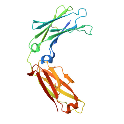
9K2Y, 9K34 - PubMed Abstract:
Enabling immunoglobulin G (IgG)-format antibodies to autonomously internalize and localize in the cytosol of targeted cells-referred to as cytosol-penetrating antibodies (cytotransmab, CT)-is challenging yet highly promising. A primary barrier to cytosolic access for CT is limited endosomal escape. Herein, we developed a second-generation (2G) CT, named in2CT4.1, featuring an endosomal acidic pH-responsive endosomal escape motif (R-W/E motif) with Arg-Trp pairs and a Glu patch in the CH3 and CL domains of IgG1/κ antibody. This motif selectively destabilizes endosomal membranes at endosomal acidic pH to facilitate cytosolic access while remaining inactive at neutral pH. The 2G CT, in2CT4.1, achieves efficient cytosolic localization at nanomolar concentrations, demonstrating approximately 3-fold higher endosomal escape efficiency compared to the first-generation CT. The potential of 2G CT is validated by engineering a cytosolic α-tubulin-targeting CT via an α-tubulin-specific variable domain in in2CT4.1. Additionally, the 2G CT effectively delivers the catalytic domain of diphtheria toxin to the cytosol of epidermal growth factor receptor-overexpressing tumor cells, resulting in near-complete suppression of tumor growth in a xenograft mouse model. These results establish 2G CT as a versatile platform for targeting cytosolic proteins and delivering therapeutic payloads, with broad potential in targeted cancer therapy and other applications.
- Department of Molecular Science and Technology, College of Engineering, Ajou University, Suwon 16499, Republic of Korea.
Organizational Affiliation: