9K29 | pdb_00009k29

Structure of the Salmonella flagellar FliPQR complex reconstituted in the peptidisc


Experimental Data Snapshot

  • Method: ELECTRON MICROSCOPY
  • Resolution: 3.00 Å
  • Aggregation State: PARTICLE 
  • Reconstruction Method: SINGLE PARTICLE 

Starting Models: experimental
View more details

wwPDB Validation   3D Report Full Report


This is version 1.1 of the entry. See complete history


Literature

A beta-cap on the FliPQR protein-export channel acts as the cap for initial flagellar rod assembly.

Kinoshita, M.Miyata, T.Makino, F.Imada, K.Namba, K.Minamino, T.

(2025) Proc Natl Acad Sci U S A 122: e2507221122-e2507221122

  • DOI: https://doi.org/10.1073/pnas.2507221122
  • Primary Citation of Related Structures:  
    9K29

  • PubMed Abstract: 

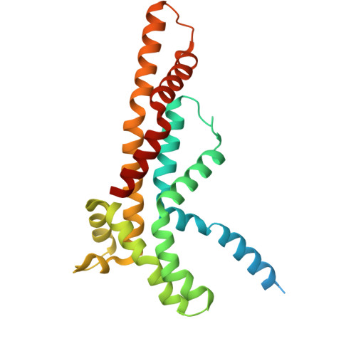
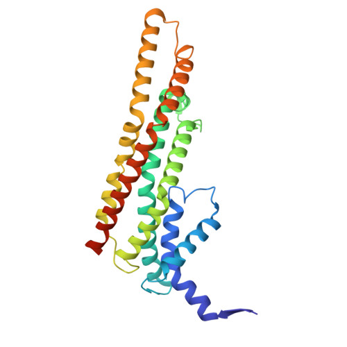
    The FliPQR complex constitutes a channel for export of the flagellar proteins involved in axial structure assembly. It also serves as a template for the assembly of the rod structure, which consists of FliE, FlgB, FlgC, FlgF, and FlgG. FliP, FliQ, and FliR assemble into a right-handed helical structure within the central pore of the flagellar basal body MS-ring, and the complex has two gates on the cytoplasmic and periplasmic sides. The periplasmic gate, formed by the N-terminal α-helices of FliP and FliR, remains closed until six FliE subunits assemble onto FliP and FliR to form the first layer of the rod, but it has remained unclear how each FliE subunit opens the gate and assembles in the absence of the rod cap required for efficient assembly of other rod proteins. Here, we present a cryoelectron microscopy structure of the FliPQR complex in closed form at 3.0 Å resolution. A β-cap, formed by the N-terminal β-strands of FliP and FliR, is located at the top of the FliPQR complex and tightly seals the closed gate. The β-cap has a narrow pore that efficiently and accurately leads the first FliE subunit to its assembly site. Interactions of FliE with FliP and FliR induce a conformational change in FliP and FliR, with their N-terminal α-helices move up and outward to open the gate. Consequently, each of the N-terminal β-strands of FliP and FliR detaches from the β-cap one after another, thereby creating a docking site for the next FliE subunit to efficiently assemble.


  • Organizational Affiliation
    • Graduate School of Frontier Biosciences, Osaka University, Suita, Osaka 565-0871, Japan.

Macromolecules
Find similar proteins by:  (by identity cutoff)  |  3D Structure
Entity ID: 1
MoleculeChains Sequence LengthOrganismDetailsImage
Flagellar biosynthetic protein FliP
A, B, C, D, E
245Salmonella enterica subsp. enterica serovar Typhimurium str. LT2Mutation(s): 0 
Gene Names: fliPflaRSTM1979
Membrane Entity: Yes 
UniProt
Find proteins for P54700 (Salmonella typhimurium (strain LT2 / SGSC1412 / ATCC 700720))
Explore P54700 
Go to UniProtKB:  P54700
Entity Groups  
Sequence Clusters30% Identity50% Identity70% Identity90% Identity95% Identity100% Identity
UniProt GroupP54700
Sequence Annotations
Expand
  • Reference Sequence
Find similar proteins by:  (by identity cutoff)  |  3D Structure
Entity ID: 2
MoleculeChains Sequence LengthOrganismDetailsImage
Flagellar biosynthetic protein FliR274Salmonella enterica subsp. enterica serovar Typhimurium str. LT2Mutation(s): 0 
Gene Names: fliRflaPSTM1981
Membrane Entity: Yes 
UniProt
Find proteins for P54702 (Salmonella typhimurium (strain LT2 / SGSC1412 / ATCC 700720))
Explore P54702 
Go to UniProtKB:  P54702
Entity Groups  
Sequence Clusters30% Identity50% Identity70% Identity90% Identity95% Identity100% Identity
UniProt GroupP54702
Sequence Annotations
Expand
  • Reference Sequence
Find similar proteins by:  (by identity cutoff)  |  3D Structure
Entity ID: 3
MoleculeChains Sequence LengthOrganismDetailsImage
Flagellar biosynthetic protein FliQ
G, H, I, J
89Salmonella enterica subsp. enterica serovar Typhimurium str. LT2Mutation(s): 0 
Gene Names: fliQflaQSTM1980
Membrane Entity: Yes 
UniProt
Find proteins for P0A1L5 (Salmonella typhimurium (strain LT2 / SGSC1412 / ATCC 700720))
Explore P0A1L5 
Go to UniProtKB:  P0A1L5
Entity Groups  
Sequence Clusters30% Identity50% Identity70% Identity90% Identity95% Identity100% Identity
UniProt GroupP0A1L5
Sequence Annotations
Expand
  • Reference Sequence
Experimental Data & Validation

Experimental Data

  • Method: ELECTRON MICROSCOPY
  • Resolution: 3.00 Å
  • Aggregation State: PARTICLE 
  • Reconstruction Method: SINGLE PARTICLE 
EM Software:
TaskSoftware PackageVersion
MODEL REFINEMENTPHENIX1.20.1_4487:

Structure Validation

View Full Validation Report


Entry History & Funding Information

Deposition Data


Funding OrganizationLocationGrant Number
Japan Society for the Promotion of Science (JSPS)JapanJP20K15749
Japan Society for the Promotion of Science (JSPS)JapanJP22K06162
Japan Society for the Promotion of Science (JSPS)JapanJP19H03182
Japan Society for the Promotion of Science (JSPS)JapanJP22H02573
Japan Society for the Promotion of Science (JSPS)JapanJP22K19274
Japan Agency for Medical Research and Development (AMED)JapanJP19am0101117
Japan Agency for Medical Research and Development (AMED)JapanJP21am0101117
Japan Agency for Medical Research and Development (AMED)JapanJP17pc0101020
Ministry of Education, Culture, Sports, Science and Technology (Japan)JapanJP20H05532
Ministry of Education, Culture, Sports, Science and Technology (Japan)JapanJP22H04844

Revision History  (Full details and data files)

  • Version 1.0: 2025-08-06
    Type: Initial release
  • Version 1.1: 2025-09-10
    Changes: Data collection, Database references