


The Crystal Structure of dsPETase05 from Biortus
Wan, T., Wang, F., Lv, Z., Lin, D., Pan, W., Shang, H., Sun, J.To be published.
Experimental Data Snapshot
Starting Model: experimental
View more details
wwPDB Validation 3D Report Full Report

Entity ID: 1 | |||||
|---|---|---|---|---|---|
| Molecule | Chains | Sequence Length | Organism | Details | Image |
| Alpha/beta hydrolase | 268 | Pseudomonadota bacterium | Mutation(s): 0 | 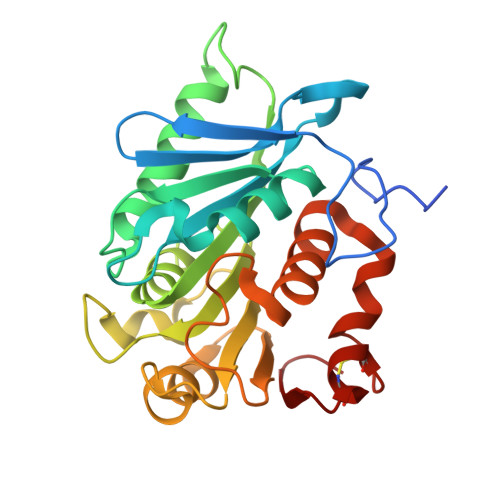 | |
UniProt | |||||
Find proteins for A0A1S8DDV9 (Halopseudomonas pachastrellae) Explore A0A1S8DDV9 Go to UniProtKB: A0A1S8DDV9 | |||||
Entity Groups | |||||
| Sequence Clusters | 30% Identity50% Identity70% Identity90% Identity95% Identity100% Identity | ||||
| UniProt Group | A0A1S8DDV9 | ||||
Sequence AnnotationsExpand | |||||
Reference Sequence | |||||
| Ligands 1 Unique | |||||
|---|---|---|---|---|---|
| ID | Chains | Name / Formula / InChI Key | 2D Diagram | 3D Interactions | |
| GOL Download:Ideal Coordinates CCD File | C [auth A], D [auth B] | GLYCEROL C3 H8 O3 PEDCQBHIVMGVHV-UHFFFAOYSA-N |  | ||
| Length ( Å ) | Angle ( ˚ ) |
|---|---|
| a = 41.066 | α = 90 |
| b = 67.273 | β = 96.529 |
| c = 85.446 | γ = 90 |
| Software Name | Purpose |
|---|---|
| REFMAC | refinement |
| XDS | data reduction |
| Aimless | data scaling |
| MoRDa | phasing |


