Structural and molecular basis for phosphate recognition by SAR11 bacteria.
Zhu, W.J., Wang, C., Liu, L., Li, J.X., Wang, H.Q., Wang, M.Q., Cao, H.Y., Chen, X.L., Qin, Q.L., Zhang, Y.Z., Sun, M.L., Wang, P.(2025) mBio 16
- PubMed: 40801535 Search on PubMed
- DOI: https://doi.org/10.1128/mbio.01654-25
- Primary Citation Related Structures:
9JWY - PubMed Abstract:
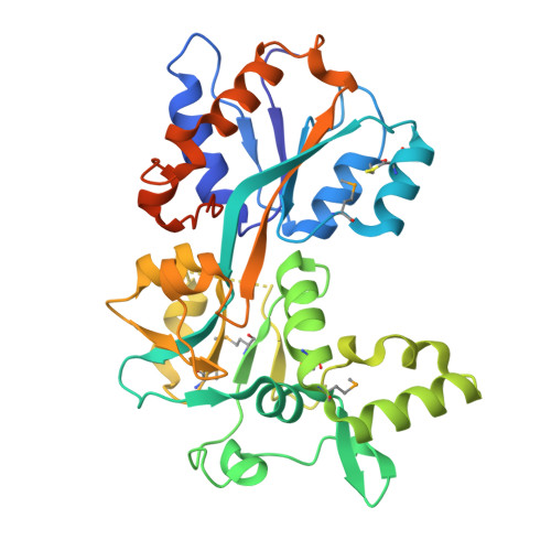
Phosphorus is a critical limiting nutrient that constrains the survival, growth, and reproduction of marine microorganisms in nutrient-limited ecosystems. Phosphorus exists in the environment in both organic and inorganic forms, with phosphate being the predominant form of inorganic phosphorus. SAR11 bacteria, a group of oligotrophic marine bacteria, possess high-affinity transporters for limiting nutrients such as phosphate, nitrogen, and organic carbon, enabling them to dominate in nutrient-depleted environments. Despite their ecological significance, the molecular mechanisms underlying phosphate transport and metabolism in SAR11 bacteria remain elusive. Here, we investigated the phosphate transport system in Candidatus Pelagibacter sp. HTCC7211, a representative SAR11 bacterium, which encodes an ATP-binding cassette-type phosphate transporter, Cp PstSCAB. We heterologously expressed and purified Cp PstS, the substrate-binding component of the transporter, and determined its crystal structure in complex with phosphate to gain insights into its substrate recognition mechanism. Microscale thermophoresis binding assays demonstrated that Cp PstS binds phosphate with high affinity, exhibiting a dissociation constant ( K d ) of 112 nM. Phylogenetic analysis placed Cp PstS in a distinct branch of previously characterized PstS proteins. Structural analysis further revealed that Cp PstS employs a unique binding site and a distinct hydrogen-bonding network for phosphate recognition and features an expanded substrate-binding cavity, suggesting potential for organic phosphorus binding. The bioinformatic analysis further indicated that Cp PstS-type phosphate-binding proteins are widely distributed among SAR11 bacteria. These findings provide valuable insights into phosphorus acquisition and utilization mechanisms in SAR11 bacteria, enhancing our understanding of their adaptation to nutrient-limited marine environments.IMPORTANCEThis study provides crucial insights into phosphate acquisition in SAR11 bacteria, a key group of oligotrophic microorganisms that thrive in nutrient-limited marine ecosystems. By characterizing the unique structural features of Cp PstS, including its distinct hydrogen-bonding network and expanded substrate-binding cavity, this research sheds light on how SAR11 bacteria adapt to limited phosphorus availability. The discovery that Cp PstS may also accommodate organic phosphorus compounds broadens our understanding of microbial nutrient acquisition. These findings have significant implications for marine biogeochemical cycles and offer new perspectives on the evolution of nutrient transport mechanisms in marine microorganisms.
- MOE Key Laboratory of Evolution and Marine Biodiversity, Frontiers Science Center for Deep Ocean Multispheres and Earth System and College of Marine Life Sciences, Ocean University of China, Qingdao, China.
Organizational Affiliation: