9JWS | pdb_00009jws

Haloquadratum walsbyi middle rhodopsin


Experimental Data Snapshot

  • Method: X-RAY DIFFRACTION
  • Resolution: 2.50 Å
  • R-Value Free: 
    0.324 (Depositor), 0.331 (DCC) 
  • R-Value Work: 
    0.296 (Depositor), 0.310 (DCC) 
  • R-Value Observed: 
    0.299 (Depositor) 

Starting Model: in silico
View more details

wwPDB Validation 3D Report Full Report

Validation slider image for 9JWS

Ligand Structure Quality Assessment 


This is version 1.1 of the entry. See complete history

Literature

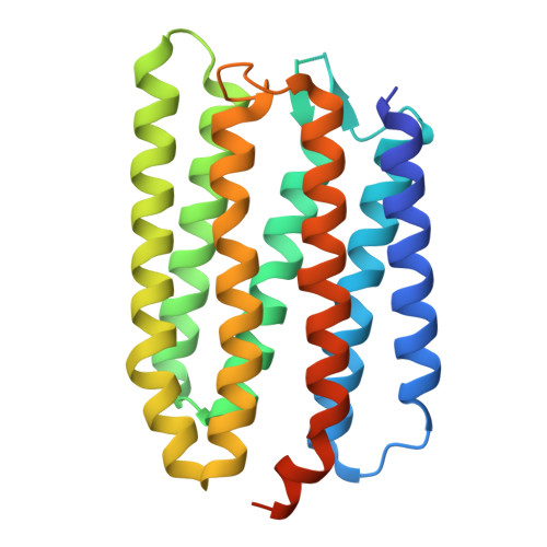
Rhodopsin from Haloquadratum walsbyi is a light-driven magnesium transporter.

Ko, L.N.Lim, G.Z.Chen, J.C.Ko, T.Li, G.Y.Yang, C.S.

(2025) Nat Commun 16: 4472-4472

  • DOI: https://doi.org/10.1038/s41467-025-59795-y
  • Primary Citation Related Structures: 
    8XHW, 9JWS

  • PubMed Abstract: 

    The functionally unknown Middle rhodopsin (HwMR) is a microbial rhodopsin (mRho) identified in Haloquadratum walsbyi, an archaeon that thrives in a 2 M MgCl 2 environment harmful to most other microorganisms. HwMR shares conserved and functionally critical residues with both bacteriorhodopsin (BR), a proton pump, and sensory rhodopsin II (SRII), which mediates phototaxis, even though HwMR exerts neither function. We previously reported HwMR as a unique mRho found to associate with Mg 2+ . Here, we show that HwMR can sense environmental Mg 2+ concentration via the D84 residue according to characteristic maximum absorption wavelength shift, photocycle kinetics, and Mg 2+ titration assay. X-ray crystallography of the wild-type HwMR and its D84N mutant produced two HwMR atomic structure models. Omit maps analysis of the wild-type HwMR model revealed D84 as a Mg 2+ binding site. On the cytoplasmic side, omit maps also revealed Mg 2+ association with T216. Both Mg 2+ sites were absent in the D84N mutant. A cell-based light-driven conductivity assay provided evidence to propose that HwMR is an inward magnesium transporter, with D84 as the primary binding site and T216 as the transportation stabilizing site. A sequential model was proposed to illustrate Mg 2+ transportation in HwMR.


  • Organizational Affiliation
    • Department of Biochemical Science and Technology, College of Life Science, National Taiwan University, Taipei, Taiwan, ROC.

Macromolecule Content 

  • Total Structure Weight: 55.67 kDa 
  • Atom Count: 3,453 
  • Modeled Residue Count: 443 
  • Deposited Residue Count: 504 
  • Unique protein chains: 1

Macromolecules

Find similar proteins by:|  3D Structure
Entity ID: 1
MoleculeChains  Sequence LengthOrganismDetailsImage
Bacteriorhodopsin-II-like protein
A, B
252Haloquadratum walsbyi DSM 16790Mutation(s): 0 
Gene Names: bop2
Membrane Entity: Yes 
UniProt
Find proteins for Q18DH5 (Haloquadratum walsbyi (strain DSM 16790 / HBSQ001))
Explore Q18DH5 
Go to UniProtKB:  Q18DH5
Entity Groups
Sequence Clusters30% Identity50% Identity70% Identity90% Identity95% Identity100% Identity
UniProt GroupQ18DH5
Sequence Annotations
Expand
Reference Sequence

Experimental Data & Validation

Experimental Data

  • Method: X-RAY DIFFRACTION
  • Resolution: 2.50 Å
  • R-Value Free:  0.324 (Depositor), 0.331 (DCC) 
  • R-Value Work:  0.296 (Depositor), 0.310 (DCC) 
  • R-Value Observed: 0.299 (Depositor) 
Space Group: P 21 21 21
Unit Cell:
Length ( Å )Angle ( ˚ )
a = 61.46α = 90
b = 62.47β = 90
c = 122.35γ = 90
Software Package:
Software NamePurpose
PHENIXrefinement
XDSdata reduction
PHENIXphasing
XSCALEdata scaling

Structure Validation

View Full Validation Report


Ligand Structure Quality Assessment 


Entry History 

& Funding Information

Deposition Data


Funding OrganizationLocationGrant Number
Not funded--

Revision History  (Full details and data files)

  • Version 1.0: 2025-05-21
    Type: Initial release
  • Version 1.1: 2025-05-28
    Changes: Database references