The C-terminal structure of the N6-methyladenosine deaminase YerA and its role in deamination.
Jia, Q., Zeng, H., Xiao, N., Tang, J., Gao, S., Xie, W.(2025) Biochem J 482
- PubMed: 39876819 Search on PubMedSearch on PubMed Central
- DOI: https://doi.org/10.1042/BCJ20240728
- Primary Citation Related Structures:
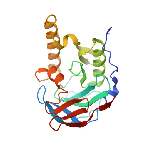
9JQL - PubMed Abstract:
The N6-methyladenine (6mA) modification is an essential epigenetic marker and plays a crucial role in processes, such as DNA repair, replication, and gene expression regulation. YerA from Bacillus subtilis is considered a novel class of enzymes that are capable of catalyzing the deamination of 6mA to produce hypoxanthine. Despite the significance of this type of enzymes in bacterial self-defense system and potential applications as a gene-editing tool, the substrate specificity, catalytic mechanism, and physiological function of YerA are currently unclear due to the lack of structural information. In the present study, we expressed the recombinant enzyme and conducted its reconstitution to yield the active form. Our deamination assays showed that N6-methyladenosine (N6-mAdo) served as a more favorable substrate than its base derivative 6mA. Here, we report the high-resolution structure of the C-terminal region of YerA, which exhibited a compact architecture composed of two antiparallel β-sheets with no obvious close structural homologs in Protein Data Bank. We also created docking models to investigate the ligand-binding pattern and found that more favorable contacts of N6-mAdo with the enzyme-binding pocket lead to its preference for N6-mAdo over 6mA. Finally, structural comparison of the N6-methyladenosine monophosphate deaminase allowed us to propose that a plausible role for this C-terminal region is to shield the active site from solvent and protect the intermediate during catalysis. Taken together, this study sheds light on the catalytic mechanism and evolutionary pathways of the promiscuous enzyme YerA, thereby contributing to our molecular understanding of epigenetic nucleoside metabolism.
- MOE Key Laboratory of Gene Function and Regulation, State Key Laboratory for Biocontrol, School of Life Sciences, Sun Yat-Sen University, Guangzhou, Guangdong 510006, People's Republic of China.
Organizational Affiliation: