Insights Into the Conformational Dynamics of the Cytoplasmic Domain of Metal-Sensing Sensor Histidine Kinase ZraS.
Mahapatra, N., Mahanta, P., Pandey, S., Acharya, R.(2025) Proteins 93: 1465-1480
- PubMed: 40062583 Search on PubMed
- DOI: https://doi.org/10.1002/prot.26819
- Primary Citation Related Structures:
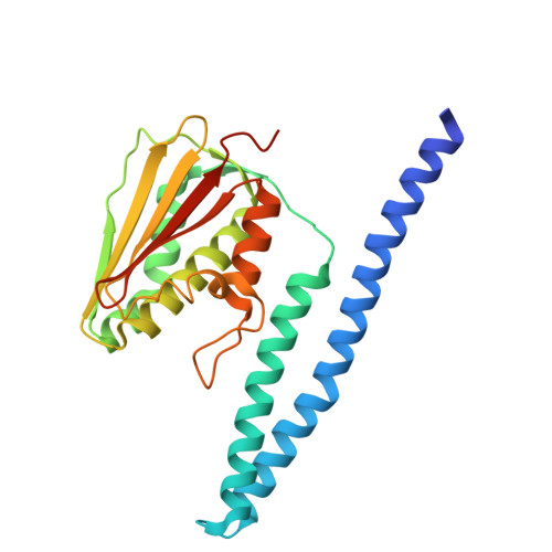
9JFJ - PubMed Abstract:
ZraS is a metal sensor integral to ZraPSR, a two-component signaling system found in enterobacters. It belongs to a family of bifunctional sensor histidine kinases (SHKs) and is speculated to sense zinc-induced stress on the bacterial envelope. Information on the structure-function relationship of sensor kinases is elusive due to the lack of full-length structures, intrinsically dynamic behavior, and difficulty trapping them in active state conformations. While the kinase domains (KDs) of a few SHKs are well characterized, they exhibit significant functional diversity attributed to their modular multi-domain arrangement in the cytoplasmic region, combined with other signal transducing elements such as simple helices, HAMP, and PAS domains. We report the crystal structure of the entire cytoplasmic region of Escherichia coli ZraS (EcZraS-CD) resolved at a resolution of 2.49 Å, comprising a unique helical linker and the KD. In the asymmetric unit, four molecules of ZraS assemble as homodimers trapped as two ligand-bound occluded conformers. Our analysis using these conformers shows that modulation of the dimer bundle through segmental helical bending, sliding, and rotation leads to the reorganization of the dimerization interface during kinase activation. Further, our analysis reveals the significance of aromatic amino acid interactions and loop residues at the dimer base in regulating the directionality of rotation during autophosphorylation. We also performed an in vitro coupled assay to determine ATPase activity. Overall, our findings provide structure-based mechanistic insights into the process of autophosphorylation in trans-acting SHKs.
- School of Biological Sciences, National Institute of Science Education and Research, Bhubaneswar, Odisha, India.
Organizational Affiliation: